Search results for: “mental health”

  • Conceptual Model Example Nursing Guide

    Conceptual Model Example Nursing Guide

    Conceptual Model Example Nursing Guide: A Comprehensive Framework for Nursing Students

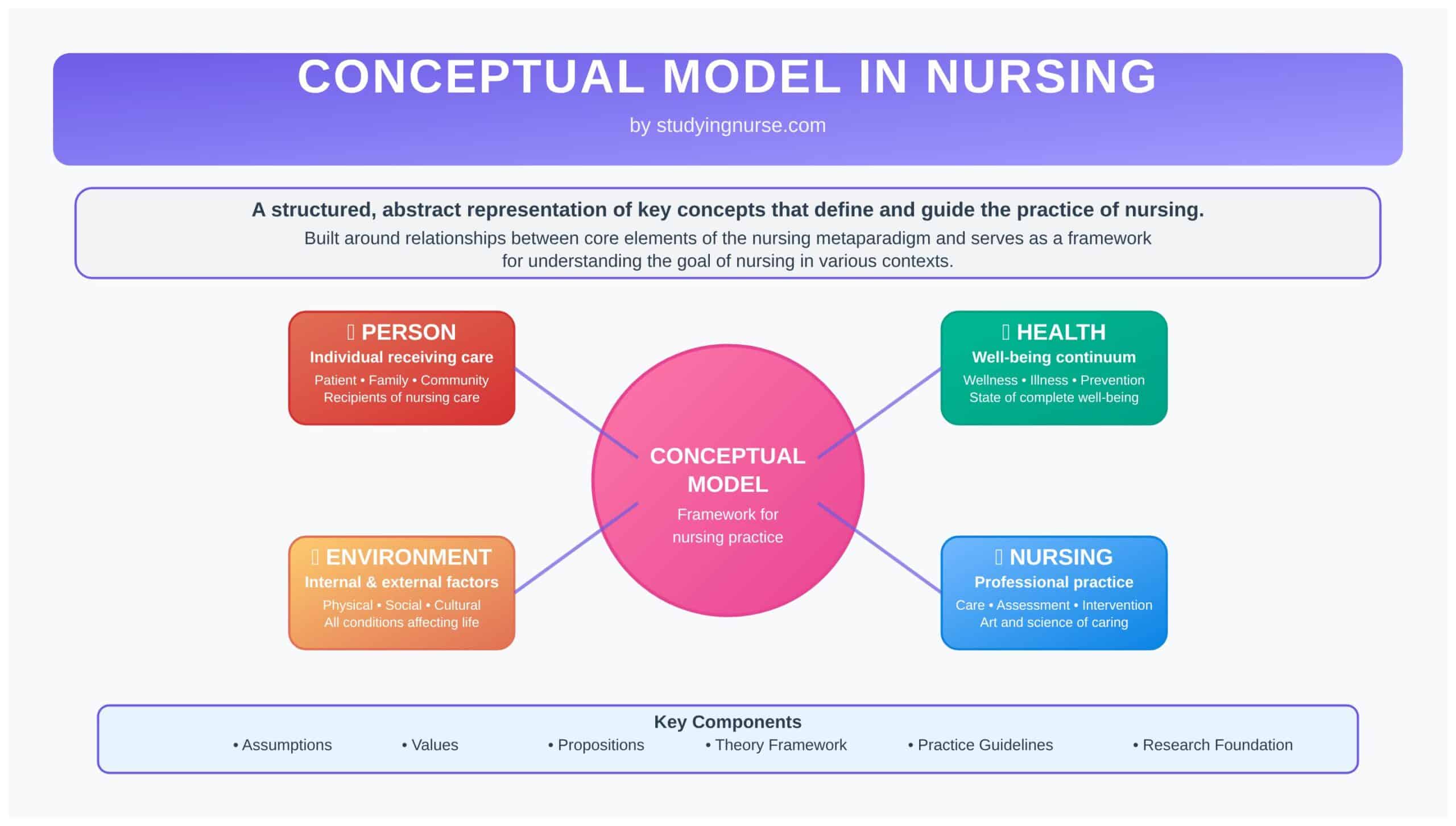
    A conceptual model in nursing is a structured, abstract representation of key concepts that define and guide the practice of nursing. These models are built around relationships between core elements of the nursing metaparadigm—person, health, environment, and nursing—and serve as a framework for understanding the goal of nursing in various contexts. A conceptual model may include assumptions, values, and propositions that inform both theory and practice.

    conceptual model in nursing
    conceptual model in nursing

    In nursing education and clinical nursing, conceptual models are used to guide nursing interventions, shape curricula, and support nursing knowledge development. They help nurses apply nursing theories in practice, offering a consistent approach to assessment, planning, and evaluation. Models like Roy’s Adaptation Model, the Neuman Systems Model, and the Behavioral System Model are commonly used to connect theoretical thinking with effective nursing care.

    These models and theories contribute to the development of nursing science by offering a basis for research, theory testing, and the improvement of care strategies. By using a conceptual model to guide decisions and actions, nurses can enhance critical thinking, ensure consistency in care, and align with evidence-based standards in the profession of nursing.

    Conceptual Model Example Nursing
    The Nursing Metaparadigm

    Understanding the Conceptual Model of Nursing

    Definition and Purpose of a Conceptual Model in Nursing

    A conceptual model in nursing is an organized and systematic representation of ideas that define the structure and content of the profession of nursing. It explains how various components of nursing—such as patients, health, environment, and care—interact and influence one another within a defined framework. Unlike specific nursing theories, which tend to focus on narrower aspects of nursing care, conceptual models provide broader guidance that links theoretical knowledge to practical application. They are often built from two or more theory concepts that help describe and explain a specific aspect of nursing.

    The purpose of a conceptual model is to serve as a guide for nursing practice, education, and research. It helps identify the goal of nursing, clarify assumptions, and outline the principles that underlie nursing actions. By offering a coherent structure, a conceptual model supports the development of nursing knowledge, the integration of theories and models, and the formation of nursing interventions that are aligned with specific health outcomes. It also serves as a foundation for nursing theory development, providing context and consistency for generating new ideas and testing them through nursing research.

    Importance of a Conceptual Framework in Nursing Practice

    A conceptual framework in nursing is essential for ensuring that care is delivered in a consistent, rational, and evidence-informed manner. It links nursing theories to clinical application, offering a structural guide for the nursing process—from assessment and diagnosis to planning, implementation, and evaluation. In clinical nursing, the use of a conceptual framework enables nurses to make informed decisions, tailor care plans, and evaluate outcomes based on theoretical understanding.

    The use of a conceptual framework is particularly relevant in complex healthcare settings, where nurses must integrate contemporary nursing knowledge with clinical judgment. For instance, in high-acuity environments, using the Neuman Systems Model can help nurses assess multiple stressors affecting a patient and implement layered defense strategies. Similarly, applying the Roy’s Adaptation Model can guide interventions that support a patient’s physiological, psychological, and sociocultural adaptation.

    Furthermore, conceptual frameworks contribute to the standardization of nursing language and practice. They enable nurses across different settings to align their care strategies, reducing variability and enhancing collaboration. As nursing models are embedded in practice, the framework also promotes accountability and critical thinking, both of which are essential in advanced nursing and professional nursing roles.

    The Nurse’s Role in Applying a Conceptual Model to Guide Care

    The nurse plays a pivotal role in interpreting and applying conceptual models to real-world scenarios. In practice, this involves selecting an appropriate model for nursing, using it to assess patient needs, and applying the model’s propositions to guide nursing interventions. The process is dynamic and often tailored to the patient population, clinical setting, and specific health conditions being addressed.

    Nurses may use the Behavioral System Model to understand patterns of behavior and design interventions that restore balance. In community health, the Conservation Model may be used to promote energy and resource conservation during illness or recovery. Through the application of these models, the nurse ensures that care is grounded in theory, culturally sensitive, and aligned with the focus of nursing practice.

    Moreover, nurses contribute to the ongoing refinement of conceptual models through nursing research and feedback from clinical outcomes. They may participate in studies that test empirical indicators for the theory concepts, thus validating or improving the models in use. Nurse researchers and educators also play a role in teaching students how to critically appraise and apply different nursing conceptual models, ensuring that the next generation of nurses continues to bridge theory and practice.

    The Nursing Metaparadigm and Its Relationship to Conceptual Models

    The nursing metaparadigm is a set of four core concepts that together define the intellectual boundaries of nursing science. These foundational elements—person, health, environment, and nursing—form the basis of all nursing conceptual models and nursing theories in practice. Each conceptual model or theory interprets these domains differently, shaping how care is delivered, how patients are assessed, and what goals are prioritized in nursing interventions.

    Understanding how a conceptual model of nursing incorporates these concepts helps nurses choose an appropriate model for their clinical context. It also ensures alignment with the broader goal of nursing, which is to promote health and well-being through theory-informed, patient-centered care.

    1. Person

    The concept of person refers to the individual receiving care, but it may also extend to families, communities, or populations, depending on the scope of the nursing model.

    • It encompasses the biological, psychological, social, spiritual, and cultural dimensions of human life.
    • In some models or theories, the person is viewed as a whole system or an energy field rather than a collection of parts.

    Examples:

    • In the Science of Unitary Human Beings (Martha Rogers), the person is seen as an indivisible energy field in constant interaction with the environment.
    • In Roy’s Adaptation Model, the person is a biopsychosocial being who adapts to internal and external stimuli through regulator and cognator mechanisms.

    This variation in interpretation influences how nurses assess needs, plan care, and evaluate patient outcomes.

    2. Health

    Health is generally defined as the degree of wellness or well-being experienced by the person. It is not merely the absence of disease but a dynamic state of balance and functioning.

    • Health can be viewed as a continuum, ranging from high-level wellness to illness or dysfunction.
    • Different conceptual models emphasize varying dimensions of health—physical, emotional, spiritual, or social.

    Examples:

    • In Neuman’s Systems Model, health is equated with system stability, and stressors disrupt that stability.
    • In the Conservation Model (Myra Levine), health is the ability to conserve energy, structural integrity, personal integrity, and social integrity.

    These perspectives help shape how nursing interventions are designed to promote, maintain, or restore health.

    3. Environment

    The environment includes all internal and external factors that affect the person. This concept is central to many nursing conceptual models, as it influences both health status and nursing responses.

    • It includes physical surroundings, social relationships, cultural influences, economic conditions, and even technological contexts.
    • Some models emphasize the reciprocal interaction between the person and environment.

    Examples:

    • In Florence Nightingale’s Environmental Theory, the physical environment (e.g., light, ventilation, cleanliness) is essential for recovery.
    • In Transcultural Nursing (Madeleine Leininger), the environment includes cultural values and practices that shape health behaviors.

    Understanding environmental influences helps nurses provide culturally competent and context-aware care.

    4. Nursing

    The concept of nursing defines the actions, attributes, and responsibilities of the nurse within the care process. It reflects how the art and science of nursing are applied to meet the needs of the person in relation to health and environment.

    • This includes direct care, advocacy, teaching, coordination, and ethical decision-making.
    • The role of nursing can be intervention-focused, adaptive, restorative, or transformational, depending on the theory or model.

    Examples:

    • In Orem’s Self-Care Deficit Theory, nursing is required when individuals cannot meet their own self-care needs.
    • In Watson’s Theory of Human Caring, nursing is seen as a healing interpersonal process that emphasizes holistic and relational care.

    These definitions affect how nurses provide a framework for care, guide clinical decisions, and articulate their contributions within healthcare teams.

    Theoretical Foundations of Nursing

    What Is a Nursing Theory?

    A nursing theory is a systematic, organized set of ideas that explains, describes, predicts, or prescribes phenomena relevant to nursing practice. These theories provide structured explanations of the art and science of nursing, guiding how nurses assess patients, plan interventions, and evaluate outcomes. At its core, a nursing theory is based on defined concepts and propositions that link back to the nursing metaparadigm—person, health, environment, and nursing.

    Nursing theories are essential to the development of nursing knowledge and the advancement of nursing science. They support evidence-based practice, contribute to theory development, and offer a foundation for nursing education and nursing research. A theory or conceptual model is often selected to align with a nurse’s clinical or academic focus, helping to unify care delivery and scholarly inquiry under a common framework.

    Examples of theories widely used in practice include Orem’s Self-Care Deficit Theory, which focuses on individuals’ ability to meet self-care needs, and Watson’s Theory of Human Caring, which centers on the relational aspects of nursing care. These existing theories demonstrate how two or more concepts can be linked to describe the focus of nursing practice in different contexts.

    Differentiating Grand Theories and Middle-Range Theories

    In nursing science, theories are commonly classified by their level of abstraction and scope of application. Two major categories—grand theories and middle-range theories—form the backbone of the theoretical foundations of nursing. Both types of theories are essential to nursing knowledge development, and both serve to guide nursing practice, inform research, and support nursing education. While they differ in scope and specificity, they share some foundational characteristics.

    Differentiating Grand Theories and Middle-Range Theories
    Differentiating Grand Theories and Middle-Range Theories

    Similarities Between Grand and Middle-Range Theories

    • Both are theory-based frameworks: They offer systematic explanations of nursing phenomena by organizing concepts and propositions relevant to the practice of nursing.
    • Grounded in the nursing metaparadigm: Each theory incorporates the core elements of the nursing metaparadigm—person, health, environment, and nursing.
    • Support the profession of nursing: Both theory types help clarify the goal of nursing, promote effective nursing care, and strengthen the scientific foundation of the discipline.
    • Used in nursing education and research: Whether broad or focused, these theories influence the design of nursing curriculum, the formation of hypotheses in nursing research, and the structuring of nursing interventions.
    • Contribute to theory development: Middle-range theories often evolve from or test grand theories, while grand theories inspire the creation of nursing conceptual models that guide practice and education.

    Differences Between Grand and Middle-Range Theories

    Scope and Abstraction

    Grand theories are broad, abstract, and philosophical.

    Middle-range theories are narrower, more specific, and practical.

    Level of Detail

    Grand theories provide a conceptual overview of nursing as a discipline.

    Middle-range theories offer detailed explanations for particular nursing phenomena (e.g., comfort, coping).

    Applicability in Clinical Practice

    Grand theories are less directly applicable in day-to-day nursing care.

    Middle-range theories are readily applicable to clinical nursing and nursing interventions.

    Origin and Development

    Grand theories are often derived from philosophical thinking and abstract reasoning.

    Middle-range theories usually evolve from empirical observation or are derived from grand theories.

    Examples

    Grand theory example: Roy’s Adaptation Model, based on a grand theory that conceptualizes individuals as adaptive systems.

    Middle-range theory example: Kolcaba’s Theory of Comfort, which offers practical strategies to enhance patient well-being.

    Testing and Research Utility

    Grand theories are not easily testable due to their abstract nature.

    Middle-range theories can be empirically tested and are often used in nursing research.

    Use in Education

    Grand theories are commonly taught in advanced nursing and theory courses to provide a broad understanding of nursing philosophy.

    Middle-range theories are used to help students apply theory to specific clinical practice situations.

    Relationship to Conceptual Models

    Grand theories often serve as the basis for nursing conceptual models (e.g., Neuman Systems Model, Science of Unitary Human Beings).

    Middle-range theories may be incorporated into or derived from these models for focused application.

    Purpose

    Grand theories aim to define the total scope and purpose of the discipline.

    Middle-range theories aim to improve specific aspects of nursing care and patient outcomes.

    Grand Theories: A Broad Philosophical Lens

    Grand nursing theories represent the most abstract level of theoretical thinking in nursing. They describe the general nature, scope, and purpose of nursing, often rooted in philosophical assumptions about human beings, health, and caring. These theories are not intended to provide immediate clinical solutions but rather to offer a conceptual framework for understanding what nursing is and why it exists.

    • Roy’s Adaptation Model, for instance, views the patient as an adaptive system that responds to internal and external stimuli. It provides a model of nursing that emphasizes adaptation as a primary goal of care.
    • Rogers’ Science of Unitary Human Beings sees individuals as irreducible energy fields, shifting the focus of nursing toward pattern recognition and environmental interaction.

    Grand theories form the foundation of nursing conceptual models and are often used in theory development, academic discourse, and conceptual analyses. They shape how nurses identify the conceptual model most appropriate for a given care philosophy or setting.

    Middle-Range Theories: Practice-Focused and Testable

    Middle-range theories emerged to address the gap between abstract theorizing and real-world nursing application. These theories are narrower in scope and more directly used to guide nursing interventions, research, and patient care. They are developed around specific phenomena such as pain management, coping, stress, or comfort, making them valuable in clinical practice and advanced nursing roles.

    • Kolcaba’s Theory of Comfort provides a clear framework for assessing and enhancing patient comfort in physical, emotional, and environmental dimensions.
    • Leininger’s Transcultural Nursing Theory focuses on culturally competent care, recognizing the role of cultural values and beliefs in health behaviors.
    • Pender’s Health Promotion Model supports proactive health behaviors and patient empowerment.

    These theories are more likely to include empirical indicators for the theory concepts, making them testable and research-driven. They also contribute significantly to nursing knowledge development by producing evidence that refines conceptual models or informs theories provide in practice.

    Conceptual Model Example Nursing
    Comparison Between Grand and Middle-Range Theories

    Connection Between Nursing Theories and Conceptual Frameworks

    The relationship between nursing theories and conceptual frameworks is foundational to theoretical thinking in the discipline. A conceptual framework serves as a broader organizing structure that helps classify and connect nursing theories. In this sense, a nursing conceptual model acts as the overarching structure, while theories provide the content that operates within it.

    • Conceptual frameworks help organize ideas, define relationships between concepts and propositions, and offer a framework for practice, education, and research.
    • Nursing theories explain specific aspects of that framework—such as adaptation, comfort, or caring—providing nursing interventions that align with the chosen model of nursing.

    For example, a nurse educator might use the Neuman Systems Model (a conceptual model) to design a curriculum, while selecting the Theory of Health Promotion (a middle-range theory) to guide teaching about preventive care strategies. In nursing research, conceptual models may be used to provide a framework for identifying variables, forming hypotheses, and interpreting data.

    This layered connection enhances clarity, consistency, and relevance in nursing practice, supporting both theory development and application.

    Conceptual Models and Theories in the Purpose of Nursing

    Both conceptual models and nursing theories are essential to articulating and advancing the purpose of nursing. They help define what nursing needed to be—historically, currently, and in the future—by framing how nurses view patients, health, and their own roles. Through their structure, these models and theories guide the development of nursing, support the formulation of nursing interventions, and influence health outcomes across different care settings.

    • Conceptual models like the Science of Unitary Human Beings or the Conservation Model emphasize the holistic nature of care, encouraging nurses to consider energy fields or resource conservation in care planning.
    • Theories provide detailed approaches to fulfilling that purpose. For instance, Peplau’s Interpersonal Relations Theory focuses on the nurse-patient relationship as the core of healing and care.

    Importantly, these theoretical foundations are not static. As contemporary nursing knowledge evolves, conceptual models and theories must be revisited and revised. This is where nurse researchers play a vital role—testing empirical indicators for the theory concepts, validating assumptions, and generating new theories through practice-based evidence.

    Together, conceptual models and theories clarify the goal of nursing, support the practice of nursing across specialties, and ensure that care is grounded in a coherent and evolving theoretical framework. They serve not only to guide individual nurses but also to unify the profession of nursing under shared philosophical and scientific principles.

    Key Conceptual Models of Nursing

    Roy’s Adaptation Model

    Developed by Sister Callista Roy, this model views the individual as a holistic adaptive system constantly responding to internal and external stimuli. The goal of nursing within this model is to promote adaptation in four adaptive modes:

    • Physiological-Physical Mode: bodily functions such as oxygenation, nutrition, and elimination.
    • Self-Concept Mode: beliefs and feelings about oneself.
    • Role Function Mode: roles the person occupies in society.
    • Interdependence Mode: relationships and support systems.

    Nursing Process in Roy’s Adaptation Model:

    Roy’s model has a six-step nursing process that mirrors the logic of the nursing process but is adapted to focus on adaptation mechanisms:

    1. Assessment of Behavior – Collect data on how the patient is currently adapting.
    2. Assessment of Stimuli – Identify focal, contextual, and residual stimuli influencing behavior.
    3. Nursing Diagnosis – Determine the ineffective or adaptive responses.
    4. Goal Setting – Establish adaptation goals for each maladaptive behavior.
    5. Intervention – Modify stimuli or strengthen adaptive processes.
    6. Evaluation – Assess whether adaptation goals have been achieved.

    This model is especially useful in rehabilitation, chronic illness management, and patient education, where continuous adaptation is essential for improving quality of life.

    Neuman Systems Model

    Developed by Betty Neuman, this model conceptualizes the client (individual, family, or community) as an open system exposed to various stressors. These stressors disrupt system stability and the nurse’s role is to help the client retain or regain system balance.

    Core Concepts:

    1. Lines of Defense:

    Flexible Line of Defense (first buffer against stressors)

    Normal Line of Defense (baseline health)

    Lines of Resistance (internal defenses activated when stressors penetrate)

    2. Stressors: Can be intrapersonal (e.g., infection), interpersonal (e.g., role conflicts), or extrapersonal (e.g., financial stress).

    Three Levels of Prevention:

    1. Primary Prevention: Reduces risk before stressor invasion (e.g., vaccinations).
    2. Secondary Prevention: Treats symptoms after stressor invasion (e.g., antibiotics for infection).
    3. Tertiary Prevention: Focuses on rehabilitation and restoring stability (e.g., therapy post-stroke).

    This model is especially effective in public health nursing, disaster response, and holistic care planning.

    Behavioral System Model

    Created by Dorothy Johnson, this model emphasizes that individuals are made up of seven subsystems, each responsible for different behaviors:

    1. Attachment/Affiliative
    2. Dependency
    3. Ingestive
    4. Eliminative
    5. Sexual
    6. Aggressive/Protective
    7. Achievement

    Each subsystem has a goal, a set of behaviors, and a choice of actions. Imbalance in any subsystem indicates illness, and the nurse acts to restore equilibrium using behavioral interventions.

    There is no fixed nursing process unique to this model, but assessment focuses on identifying maladaptive behaviors and their external/internal causes, followed by interventions to re-establish stability in the affected subsystem.

    Example: A patient with a poor nutritional intake may be experiencing a disturbance in the ingestive subsystem; the nurse would assess triggers (e.g., pain, anxiety) and intervene accordingly.

    Conservation Model

    Developed by Myra Levine, this model centers on promoting adaptation and maintaining integrity through conservation of:

    • Energy – supporting rest and nutrition
    • Structural Integrity – maintaining or restoring physical body structures
    • Personal Integrity – respecting individual dignity and self-worth
    • Social Integrity – sustaining relationships and roles

    The nurse’s role is to minimize the impact of illness while promoting healing through conservation. This is especially applicable in acute care, surgical recovery, and geriatric nursing, where physical and emotional reserves must be preserved.

    This model complements the traditional nursing process, with a focus on individualized care and assessing for threats to any of the four conservation principles. For instance, after surgery, interventions might aim to conserve energy and structural integrity through controlled activity and wound care.

    Science of Unitary Human Beings

    Formulated by Martha Rogers, this highly abstract model redefines the human as an energy field in continuous interaction with the environment. The goal of nursing is to promote harmony between these fields through patterning activities.

    Key Principles:

    Integrality: The continuous mutual interaction of person and environment.

    Resonancy: The change in human and environmental fields over time.

    Helicity: The unpredictable but increasing diversity and complexity of human patterns.

    Rather than traditional assessments, this model encourages the nurse to focus on non-invasive interventions such as guided imagery, presence, music therapy, or touch to influence energy patterns.

    It is widely used in holistic nursing, palliative care, and alternative/complementary medicine, and serves as a foundation for innovative, theory-guided nursing care.

    Transcultural Nursing Model

    Pioneered by Madeleine Leininger, this model focuses on providing culturally competent care by understanding patients’ worldviews, cultural values, and health beliefs.

    Key Concepts:

    Cultural Care Preservation – supporting beneficial cultural practices.

    Cultural Care Accommodation – adapting care to align with cultural needs.

    Cultural Care Repatterning – helping patients modify harmful practices.

    Leininger developed the Sunrise Model, a visual framework to assess cultural dimensions like technological factors, religious beliefs, kinship structures, politics, and economics—all of which influence health and care decisions.

    In practical terms, a nurse using this model might:

    • Use interpreters for non-English speakers
    • Respect fasting practices during Ramadan
    • Integrate traditional healing rituals when safe

    This model is essential in today’s multicultural clinical environments, supporting culturally safe, respectful, and effective patient-centered care.

    Conceptual Model Example Nursing
    Key Conceptual Models of Nursing

    Application of Conceptual Models in Nursing Education and Practice

    Integrating Nursing Conceptual Models into the Curriculum

    Integrating a nursing conceptual model into curriculum design ensures that nursing programs preserve the unique framework and philosophy of nursing, rather than defaulting to a purely biomedical approach. Frameworks like Roy’s Adaptation Model, Neuman Systems Model, or Leininger’s Transcultural Nursing Model provide a conceptual context for organizing content around the nursing metaparadigm and the purpose of nursing 


    At the graduate level—especially for Nurse Practitioner or Advanced Practice Nurse tracks—using a nursing model helps differentiate advanced nursing roles. It fosters a professional identity rooted in nursing knowledge and caring, distinct from purely clinical or technical function 

    Enhancing Theoretical Knowledge Through Conceptual Frameworks

    A conceptual framework provides structure to nursing theory by connecting abstract concepts and nursing theories to practical application and research. In education, frameworks help students engage with theory, identify variables, and connect nursing theory to real patient care scenarios. This approach aids in theory development, empirical testing, and research design.


    Framework-based teaching encourages integration of nursing knowledge development through case studies, assignments, and simulated practice rooted in theory. For example, using Orem’s Self‑Care Theory in coursework encourages students to design plans promoting patient self‑efficacy. Using Pender’s Health Promotion Model helps educators illustrate how behavior-specific cognitions influence health outcomes

    Use of Conceptual Models in Clinical Nursing Practice

    Conceptual models are not just academic; they shape clinical nursing care directly. Fawcett describes a reciprocal relationship between theory and practice, where explicit models define standards, focus areas, and content for the nursing process—and clinical practice, in turn, validates or refines these models 


    In practice, conceptual models like Roy’s Adaptation Model help nurses design care plans that promote adaptation to illness; Neuman’s Systems Model supports holistic stress assessment and preventive care; Leininger’s Transcultural Model ensures culturally congruent care delivery

    Guiding the Nursing Process with Conceptual Frameworks

    Conceptual frameworks offer structure for the nursing process—assessment, diagnosis, planning, intervention, and evaluation—by clarifying priorities and relationships.

    Within Roy’s Adaptation Model, the care process explicitly examines stimuli and behaviors, plans adaptation goals, delivers interventions, and evaluates adaptation outcomes.

    Under the Neuman Systems Model, nurses assess environmental stressors and system defenses, select prevention levels, implement care, and monitor system stability.
    These models add coherence and consistency to nursing assessment and decision-making across clinical settings

    Applying Conceptual Models to Nursing Interventions

    Applying a conceptual model to guide nursing interventions ensures alignment of care with theory, improving both structure and outcomes.

    1. Identify a relevant model (e.g., Roy’s Adaptation, Orem’s Self‑Care, Leininger’s Culture Care).
    2. Assess patient needs using model-specific concepts (e.g., identifying adaptation behaviors or cultural beliefs).
    3. Design care plans with targeted nursing interventions rooted in the theory (e.g., stimuli modification, comfort strategies, cultural alignment).
    4. Implement evidence-based practices based on the model’s propositions. For instance, Roy’s model informs interventions to adjust focal stimuli, while Neuman’s guides preventive care strategies.
    5. Evaluate measurable outcomes and refine care plans. Use reflective practice and feedback to refine how the model informs care and contributes to nursing knowledge development

    Nursing Theory Development and Research

    Advancing Nursing Knowledge Through Theory Development

    Theory development lies at the heart of expanding nursing knowledge and advancing nursing science. When nurse scholars identify gaps in practice or emerging health challenges, they craft new theories or extend existing ones by defining:

    Concepts (e.g. adaptation, comfort, self-care)

    Definitions clarifying the concepts

    Relationships among those concepts

    Underlying assumptions that guide the theoretical framework 

    This structured process enables a conceptual framework that forms the backbone of nursing conceptual models. As healthcare environments evolve—due to chronic disease prevalence, demographic shifts, or technological changes—developing new or revised theories becomes essential. Such theory development helps nurses provide effective nursing care, refine nursing interventions, and align with contemporary needs in patient care

    The Role of Nurse Researchers in Conceptual and Theoretical Growth

    Nurse researchers are pivotal in driving theory development and conceptual growth within the discipline. Drawing on clinical experience, academic inquiry, and patient data, they:

    • Identify deficiencies where existing theories may be insufficient.
    • Develop or customize nursing models or theories to address real-world problems.
    • Publish and disseminate findings through conferences, journals, and teaching 

    Nurse researchers also engage in interdisciplinary collaborations, integrating insights from psychology, sociology, and public health. This enriches theory-building and supports the creation of middle‑range theories that are both grounded in evidence and sensitive to practice contexts

    Empirical Indicators and Validation of Nursing Models

    A crucial component of theory development is empirical validation—using measurable indicators to test whether theoretical propositions hold true in practice. Empirical indicators include:

    • Instruments (e.g., questionnaires)
    • Assessment tools
    • Interventions specifically designed to operationalize theoretical concepts.

    For example, a comfort scale aligned with Kolcaba’s Theory of Comfort may measure patient-reported comfort levels across dimensions of physical, emotional, and environmental well-being. The nurse researcher can then correlate these data with interventions to support theory validation. Construct validity is tested through methods such as factor analysis, structural equation modeling, or multitrait–multimethod approaches to ensure indicators accurately represent the concepts they measure

    Challenges in Nursing Theory Development

    Despite its importance, nursing theory development faces several intertwined challenges:

    1. Philosophical inconsistency
      Nursing draws from diverse philosophical traditions (positivism, historicism, holistic frameworks), leading to fragmentation in theoretical foundations. This undermines coherence in models and theories across the profession 
    2. Lack of empirical validation
      Many theories remain untested or inadequately validated in clinical settings. Without empirical support, theoretical relationships may fail under practical conditions, limiting usability and trust among practitioners 
    3. Theory–practice gap
      Abstract theoretical language and complex constructs can make it difficult for clinical nursing staff to translate models into everyday care. Many nurses report that theories are too academic and impractical for bedside use, widening the gap between scholarly knowledge and routine practice 
    4. Multiplicity of competing paradigms
      Nursing lacks a universally accepted unified theory. Instead, a plurality of models and theories exist, which can create confusion and inconsistent practice across settings and practitioners 
    5. Challenges with modification and evolution
      Some theories are rigid and resist adaptation, which hinders their evolution in light of new evidence or changing care contexts. The inability to refine existing theories limits progress in theory development and theoretical growth

    Frequently Asked Questions

    What are examples of conceptual models in nursing?

    Examples include:

    Roy’s Adaptation Model – views the patient as an adaptive system.

    Neuman Systems Model – focuses on stress and the patient’s response.

    Orem’s Self-Care Deficit Theory – emphasizes self-care needs and abilities.

    Watson’s Theory of Human Caring – centers on holistic caring and the nurse–patient relationship.

    King’s Goal Attainment Theory – highlights goal-setting between nurse and patient.

    Leininger’s Transcultural Nursing Model – integrates cultural awareness in nursing care.

    What are some examples of a conceptual model?


    Outside of nursing, conceptual models include:

    Maslow’s Hierarchy of Needs – a psychological model of human motivation.

    Health Belief Model – used in public health to explain health behaviors.
    In nursing, examples include:

    The Science of Unitary Human Beings (Martha Rogers)

    Johnson’s Behavioral System Model

    What are the 4 key concepts in nursing models?


    The four universal concepts, known as the nursing metaparadigm, are:

    Person – the recipient of nursing care (individual, family, or community).

    Health – the level of wellness or well-being.

    Environment – the internal and external factors affecting the person.

    Nursing – the actions, roles, and functions of the nurse.

    What are the concepts of the nursing model?


    Nursing models include both the metaparadigm concepts (person, health, environment, nursing) and model-specific concepts depending on the theory. For example:

    In Roy’s model, key concepts include adaptation, stimuli, and coping mechanisms.

    In Orem’s model, the concepts are self-care, self-care deficit, and nursing systems.
    These guide how care is planned, delivered, and evaluated.
  • Anselmo Lopez iHuman Case Study and Best Guide 2025

    Anselmo Lopez iHuman Case Study and Best Guide 2025

    Anselmo Lopez iHuman Case Study

    Anselmo Lopez is a 57-year-old Hispanic male construction/flooring worker presenting to the clinic with elevated blood pressure readings of approximately 170/92 discovered at a community wellness event. He reports a history of self-treating acid reflux with over-the-counter omeprazole and bilateral knee pain with ibuprofen. The patient has been absent from preventative health screenings for several years and consumes elevated processed foods high in fat and sodium.

    In this comprehensive guide, we’ll walk you through how to approach his case, from initial cardiovascular assessment through systematic physical examination to the final diagnosis of Essential Hypertension. You’ll learn the key clinical reasoning steps for hypertensive disorders, what the iHuman grading rubric expects, and a complete step-by-step solution to help you confidently navigate this essential primary care case simulation involving hypertension diagnosis and management.

    Anselmo Lopez iHuman
    Anselmo Lopez iHuman Case Study Guide

    Anselmo Lopez iHuman Case Overview (Doorway Information)

    Patient Overview: Anselmo Lopez is a 57-year-old Hispanic male presenting with chief complaint of elevated blood pressure discovered at a community wellness event with readings of 172/94 left arm and 178/98 right arm. He was brought to the clinic for comprehensive evaluation and management of newly discovered hypertension.

    Key Background Information:

    • Age/Gender: 57-year-old Hispanic male
    • Height/Weight: 195 lbs (BMI: 28.8) – overweight
    • Occupation: Construction/flooring worker
    • Chief Complaint: Elevated blood pressure readings at health fair
    • Blood Pressure: 172/94 mmHg (left arm), 178/98 mmHg (right arm)
    • Presenting Situation: Adult primary care clinic visit for hypertension evaluation
    • Associated Conditions: Acid reflux, bilateral knee pain
    • Significant History: ACL repair (unknown date), family history of hypertension and diabetes
    • Current Status: Previously undiagnosed hypertensive adult male
    • Risk Factors: Age, ethnicity, obesity, poor diet, sedentary occupation
    • Physical Findings: Prominent AV nicking (3:1), laterally displaced PMI

    The patient appears well but has been avoiding medical care for several years. His presentation with significantly elevated blood pressure, retinal changes, and cardiac findings suggests established hypertension requiring immediate intervention. This makes it an excellent case for learning systematic adult cardiovascular assessment and evidence-based hypertension management principles.

    Anselmo Lopez (57 y/o Hispanic male) – Adult Cardiovascular Assessment

    • CC: Elevated blood pressure readings at community health fair
    • MSAP: Essential Hypertension – requiring antihypertensive therapy and lifestyle modifications
    • Associated symptoms: Occasional headaches, fatigue, bilateral knee pain
    • History: Acid reflux on omeprazole, knee pain on NSAIDs, previous ACL repair
    • High-risk factors: Age 57, Hispanic ethnicity, obesity, poor diet, family history

    History Questions:

    • How can I help you today?
    • What brought you to the clinic today?
    • Tell me about your blood pressure readings at the health fair?
    • Have you ever been told you have high blood pressure before?
    • When was your last visit to a healthcare provider?
    • Do you take any medications regularly?
    • Do you have any chest pain or shortness of breath?
    • Do you experience headaches or dizziness?
    • Tell me about your work – what do you do for a living?
    • Do you exercise regularly?
    • Describe your typical diet for me?
    • How much salt would you say you consume daily?
    • Do you smoke or use tobacco products?
    • How much alcohol do you drink per week?
    • Do you have trouble sleeping or snore loudly?
    • Any family history of heart disease or high blood pressure?
    • Have you had any surgeries in the past?
    • Do you have any ongoing medical problems?
    • Are you taking any over-the-counter medications?
    • Any allergies to medications?
    • Do you have any concerns about your health?

    Physical Exam:

    Vitals: Blood pressure (both arms), pulse, respirations, temperature, weight, height, BMI calculation

    General appearance: Level of distress, overall appearance, body habitus, apparent state of health

    Cardiovascular Assessment:

    • Comprehensive blood pressure measurement in both arms
    • Heart rate and rhythm evaluation
    • Point of maximal impulse location and character
    • Assessment for murmurs, gallops, or extra heart sounds
    • Peripheral pulse examination (radial, femoral, pedal pulses)
    • Assessment for jugular venous distension
    • Evaluation for peripheral edema

    Fundoscopic Examination:

    • Detailed retinal examination for hypertensive retinopathy
    • Assessment for arteriovenous nicking, cotton wool spots, flame hemorrhages
    • Evaluation of cup-to-disc ratio and papilledema

    Neck Assessment:

    • Carotid pulse examination and auscultation for bruits
    • Thyroid gland palpation
    • Assessment of jugular venous pressure

    Respiratory Assessment:

    • Lung examination for adventitious sounds
    • Assessment of respiratory effort and pattern
    • Evaluation for signs of pulmonary edema

    Abdominal Assessment:

    • Auscultation for renal artery bruits
    • Palpation for abdominal aortic aneurysm
    • Assessment of liver size and tenderness
    • Evaluation for masses or organomegaly

    Neurological Assessment:

    • Assessment of mental status and orientation
    • Evaluation for focal neurological deficits
    • Assessment of reflexes and motor function

    Assessment Note:

    A.L. is a 57-year-old Hispanic male presenting with newly discovered essential hypertension. Physical examination notable for significantly elevated blood pressure readings (172/94 and 178/98), evidence of target organ damage with prominent arteriovenous nicking (3:1) on fundoscopic examination, and laterally displaced point of maximal impulse suggesting left ventricular hypertrophy. His presentation with multiple cardiovascular risk factors and evidence of chronic hypertensive changes indicates established disease requiring comprehensive management.

    Diagnostic Testing: Basic metabolic panel, lipid profile, urinalysis with microscopy, electrocardiogram, echocardiogram, chest X-ray, thyroid function tests

    Diagnosis: Essential Hypertension with evidence of target organ damage

    Plan:

    • Initiate antihypertensive therapy with ACE inhibitor or thiazide diuretic
    • Comprehensive lifestyle modifications including dietary changes and exercise
    • Patient education about hypertension management and medication compliance
    • Regular blood pressure monitoring and follow-up care
    • Screening for additional cardiovascular risk factors
    • Referral for ophthalmologic evaluation of retinal changes

    Anselmo Lopez SOAP Note

    Patient: Anselmo Lopez

    Subjective Data

    CC: 57-year-old Hispanic male presents with “high blood pressure found at health fair”

    HPI: 57-year-old previously undiagnosed male presents to primary care clinic for evaluation of elevated blood pressure discovered at a community wellness event. Patient reports blood pressure readings of approximately 170/92 taken at the health fair, which prompted recommendation for immediate medical evaluation. He admits to avoiding healthcare for “several years” and reports he has been told in the past that his blood pressure was elevated but never sought treatment. Patient denies chest pain, shortness of breath, or palpitations but reports occasional headaches and generalized fatigue that he attributes to work stress. He describes his occupation as construction and flooring work, which involves heavy physical labor. Patient acknowledges poor dietary habits with frequent consumption of processed foods, fast food, and high sodium intake. He denies current tobacco use but reports social alcohol consumption on weekends.

    Medications: Omeprazole 20mg daily (OTC) for acid reflux, Ibuprofen 400mg as needed for bilateral knee pain

    Allergies: No known drug, food, or environmental allergies reported

    PMH: History of acid reflux managed with OTC omeprazole, bilateral knee pain treated with NSAIDs, surgical history significant for ACL repair (unknown date), no previous diagnoses of hypertension or cardiac disease, absent from preventative healthcare for multiple years

    Family History: Significant for hypertension and diabetes mellitus in family members

    Social History: 57-year-old construction worker, poor dietary habits with consumption of processed foods high in fat and sodium, occasional social alcohol use, denies tobacco or illicit drug use, lives independently, works full-time in physically demanding job

    Review of Systems: Notable for occasional headaches, generalized fatigue, bilateral knee pain, and symptoms of acid reflux. Denies chest pain, shortness of breath, palpitations, syncope, vision changes, nausea, vomiting, or neurological symptoms.

    Objective Data

    General: Alert, oriented 57-year-old Hispanic male in no acute distress, appears stated age, overweight body habitus

    Vital Signs: Blood Pressure 172/94 mmHg (left arm), 178/98 mmHg (right arm), Heart Rate 82 bpm, Respiratory Rate 16 breaths/min, Temperature 98.6°F, Weight 195 lbs, BMI 28.8 (overweight)

    Physical Examination:

    • Appearance: Alert and oriented, cooperative, no acute distress, overweight male
    • HEENT: Normocephalic, atraumatic, fundoscopic examination reveals prominent arteriovenous nicking (3:1), consistent with hypertensive retinopathy, no papilledema, normal tympanic membranes
    • Neck: No thyromegaly, no lymphadenopathy, carotid pulses 2+ without bruits, no jugular venous distension
    • Cardiovascular: Point of maximal impulse laterally displaced, suggesting left ventricular hypertrophy, regular rate and rhythm, no murmurs, gallops, or rubs, peripheral pulses 2+ throughout
    • Respiratory: Chest expansion symmetrical, lungs clear to auscultation bilaterally, no accessory muscle use, no signs of pulmonary edema
    • Abdomen: Soft, non-tender, normoactive bowel sounds, no hepatosplenomegaly, no palpable masses, no abdominal bruits
    • Extremities: No pedal edema, normal range of motion, bilateral knee tenderness with palpation
    • Neurologic: Alert and oriented x4, no focal neurological deficits, normal reflexes, normal gait

    Assessment

    Primary Diagnosis: Essential Hypertension with evidence of target organ damage (I10)

    Secondary Concerns:

    • Hypertensive retinopathy
    • Possible left ventricular hypertrophy
    • Obesity (BMI 28.8)
    • Cardiovascular risk factor modification needed

    Differential Diagnoses

    Essential Hypertension: Most likely diagnosis given patient’s age, ethnicity, family history, and lifestyle factors. The presence of target organ damage including retinal arteriovenous nicking and laterally displaced PMI suggests chronic, established hypertension. The constellation of risk factors including age over 55, Hispanic ethnicity, obesity, poor dietary habits, and positive family history strongly supports primary hypertension.

    Secondary Hypertension: Less likely given the typical presentation and risk factor profile. However, given the significantly elevated readings, secondary causes should be considered including renal artery stenosis, primary aldosteronism, pheochromocytoma, or thyroid disorders. The absence of specific symptoms or physical findings makes secondary causes less probable.

    White Coat Hypertension: Possible consideration given the clinical setting; however, the presence of target organ damage (retinal changes and cardiac findings) suggests true sustained hypertension rather than situational elevation.

    Renovascular Hypertension: Could be considered given the severity of presentation, but absence of abdominal bruits and typical demographic make this less likely. Further evaluation with renal function assessment would be appropriate.

    Sleep Apnea-Related Hypertension: Patient is at risk for obstructive sleep apnea due to age, gender, and hypertension. The combination of obesity and hypertension raises suspicion for sleep-disordered breathing as a contributing factor.

    Most Likely Diagnosis: Essential Hypertension evidenced by significantly elevated blood pressure readings, evidence of chronic target organ damage, multiple cardiovascular risk factors, and positive family history in appropriate demographic population.

    Plan

    Health Promotion: ✓ Lifestyle Modifications – Comprehensive dietary counseling focusing on DASH diet principles, sodium restriction (<2300mg daily), weight reduction strategies ✓ Exercise Prescription – Structured aerobic exercise program 150 minutes moderate intensity weekly, considering occupational physical demands ✓ Cardiovascular Risk Reduction – Smoking cessation counseling if applicable, alcohol moderation, stress management techniques ✓ Patient Education – Understanding of hypertension, importance of medication compliance, home blood pressure monitoring

    Screening: ✓ Cardiovascular Risk Assessment – Lipid profile, diabetes screening, electrocardiogram, echocardiogram to assess left ventricular function ✓ Target Organ Damage Evaluation – Urinalysis with microalbumin, serum creatinine, ophthalmologic referral for comprehensive retinal evaluation ✓ Secondary Hypertension Screening – Basic metabolic panel, thyroid function tests, consideration of sleep study

    Interventions: ✓ Antihypertensive Therapy – Initiate ACE inhibitor (Lisinopril 10mg daily) or thiazide diuretic (Hydrochlorothiazide 25mg daily) based on patient characteristics and contraindications ✓ Medication Management – Review current NSAID use for knee pain, consider alternatives due to hypertensive effects ✓ Monitoring Plan – Return visit in 2-4 weeks for blood pressure recheck and medication tolerance assessment ✓ Long-term Management – Goal blood pressure <130/80 mmHg, regular follow-up every 3-6 months once stable

    Anselmo Lopez iHuman
    Anselmo Lopez SOAP Note

    Complete Step-by-Step Guide to Writing the Anselmo Lopez iHuman Case Study

    Completing the Anselmo Lopez iHuman case requires a systematic approach focused on adult cardiovascular assessment and evidence-based hypertension management. This comprehensive guide will walk you through each section of the simulation, providing specific strategies and key points to ensure you achieve the required 70% score.

    Step 1: Pre-Case Preparation and Initial Approach

    Before diving into the case, review the doorway information and formulate your initial clinical approach.

    Key Information to Note:

    • 57-year-old Hispanic male with newly discovered hypertension
    • Significantly elevated blood pressure readings from health fair
    • Multiple cardiovascular risk factors present
    • Evidence of target organ damage on examination

    Initial Clinical Mindset: Approach this case with essential hypertension as your primary consideration. The age (57 years old), ethnicity (Hispanic), occupational factors, and family history immediately suggest primary hypertension, which accounts for 90-95% of all hypertensive cases in adults.

    Step 2: Conducting the History of Present Illness (HPI)

    The HPI is crucial for establishing the foundation of your cardiovascular diagnosis. Use the OLDCARTS mnemonic systematically:

    Onset: Ask about when elevated blood pressure was first discovered

    • Key points to elicit: Health fair screening, previous elevated readings, duration of symptoms

    Location: Determine specific areas of concern or symptoms

    • Target response: Headache location, chest discomfort, visual changes

    Duration: How long patient has been avoiding medical care

    • Important detail: Several years without preventative care or blood pressure monitoring

    Character: Detailed description of any associated symptoms

    • Critical descriptors: Headache quality, fatigue patterns, exercise tolerance

    Aggravating factors: What makes symptoms worse

    • Essential findings: Work stress, dietary factors, physical exertion

    Relieving factors: What provides symptom relief

    • Key response: Rest, changes in position, OTC medications

    Timing: Pattern of symptom variation throughout day

    • Important pattern: Morning headaches, fatigue patterns, work-related symptoms

    Severity: Impact on patient’s daily function and work capacity

    • Typical response: Functional impairment, work performance, quality of life

    Step 3: Review of Systems (ROS)

    Conduct a thorough but focused ROS, paying special attention to cardiovascular and related systems:

    Constitutional:

    • Fatigue, weakness, weight changes
    • Sleep quality and potential sleep apnea symptoms
    • Exercise tolerance and energy levels

    Cardiovascular:

    • Chest pain, palpitations, shortness of breath
    • Peripheral edema, orthopnea, paroxysmal nocturnal dyspnea
    • Claudication or peripheral vascular symptoms

    Neurological:

    • Headaches, dizziness, visual changes
    • Weakness, numbness, cognitive changes
    • History of stroke or transient ischemic attacks

    Genitourinary:

    • Urinary frequency, nocturia, hematuria
    • Kidney disease symptoms or history
    • Sexual dysfunction (medication-related)

    Step 4: Past Medical History, Social History, and Family History

    Past Medical History:

    • Previous blood pressure readings or diagnoses
    • Cardiovascular disease, stroke, kidney disease
    • Diabetes mellitus, thyroid disorders
    • Medication history and responses

    Social History:

    • Occupational exposures and physical demands
    • Dietary patterns, sodium intake, alcohol consumption
    • Exercise habits and lifestyle factors
    • Support systems and healthcare access

    Family History:

    • Family history of hypertension and diabetes mellitus
    • Premature cardiovascular disease in relatives
    • Stroke, kidney disease, or sudden cardiac death

    Step 5: Physical Examination Strategy

    Perform a comprehensive cardiovascular physical examination:

    General Assessment:

    • Expected findings: Well-appearing overweight male, no acute distress
    • Critical component: Overall cardiovascular risk assessment

    Vital Signs:

    • Look for: Significantly elevated blood pressure in both arms, normal heart rate and respiratory rate

    Cardiovascular Examination:

    • Comprehensive cardiac assessment: laterally displaced PMI suggesting left ventricular hypertrophy
    • Look for: Murmurs, gallops, peripheral pulses, signs of heart failure

    Fundoscopic Examination:

    • Detailed retinal assessment: prominent arteriovenous nicking (3:1)
    • Look for: Cotton wool spots, flame hemorrhages, papilledema

    Systemic Examination:

    • Abdominal examination for bruits and organomegaly
    • Extremity examination for edema and peripheral pulses
    • Neurological assessment for end-organ damage

    Step 6: Developing Differential Diagnoses

    Propose at least 4-5 appropriate differentials with rationales:

    Primary Consideration: Essential Hypertension

    • Supporting evidence: Age, ethnicity, family history, lifestyle factors, target organ damage

    Secondary Considerations:

    • Secondary hypertension: Renal, endocrine, or vascular causes
    • White coat hypertension: Less likely given target organ damage
    • Sleep apnea-related hypertension: Obesity and demographic factors
    • Medication-induced hypertension: NSAIDs for knee pain

    Step 7: Diagnostic Test Interpretation

    Interpret clinical findings to support your diagnosis:

    Expected Key Findings:

    • Electrocardiogram showing left ventricular hypertrophy
    • Elevated serum creatinine or proteinuria indicating kidney involvement
    • Normal or elevated glucose levels
    • Dyslipidemia on lipid panel

    Step 8: Final Diagnosis and Most Significant Active Problem (MSAP)

    Primary Diagnosis: Essential Hypertension with target organ damage

    Justification:

    • Significantly elevated blood pressure readings (172/94, 178/98)
    • Evidence of chronic hypertensive changes (retinal and cardiac)
    • Multiple cardiovascular risk factors present
    • Typical demographic and clinical presentation

    MSAP Selection: Choose “Essential Hypertension” as your Most Significant Active Problem, as this represents the primary cardiovascular condition requiring immediate intervention and long-term management.

    Step 9: Comprehensive Management Plan

    Develop a multi-faceted treatment approach:

    Immediate Management:

    • Antihypertensive therapy initiation (ACE inhibitor or thiazide diuretic)
    • Lifestyle modification counseling
    • Risk factor assessment and modification

    Short-term Management:

    • Medication regimen: Lisinopril 10mg daily or HCTZ 25mg daily
    • Dietary counseling: DASH diet, sodium restriction (<2300mg daily)
    • Exercise prescription: 150 minutes moderate aerobic activity weekly

    Long-term Management:

    • Target blood pressure <130/80 mmHg
    • Regular follow-up every 3-6 months once stable
    • Annual cardiovascular risk assessment
    • Screening for complications and comorbidities

    Patient Education:

    • Understanding hypertension as “silent killer”
    • Importance of medication compliance and lifestyle changes
    • Home blood pressure monitoring techniques
    • When to seek immediate medical attention

    Step 10: Documentation and Submission Tips

    Writing Your Summary:

    • Create a concise 350-word summary explaining your clinical reasoning for hypertension diagnosis
    • Include how you arrived at the problem list with consideration of cardiovascular risk factors
    • Cite specific assessment findings and evidence of target organ damage
    • Use professional cardiovascular terminology

    Key Documentation Elements:

    • Assessment Statement: Brief patient summary with key cardiovascular findings
    • Clinical Reasoning: Explain diagnostic thought process for essential hypertension
    • Evidence-Based Management: Link treatment choices to current hypertension guidelines
    • Risk Stratification: Address cardiovascular risk factors and prevention strategies

    Final Submission Checklist:

    • ✓ Complete cardiovascular history with systematic symptom assessment
    • ✓ Comprehensive physical examination including detailed cardiac and fundoscopic assessment
    • ✓ Appropriate differential diagnoses with cardiovascular considerations
    • ✓ Correct final diagnosis and MSAP
    • ✓ Evidence-based antihypertensive management plan
    • ✓ Professional documentation with proper cardiovascular terminology

    Anselmo Lopez iHuman Case Summary Grading Criteria

    The Anselmo Lopez iHuman case will evaluate you across several critical domains to ensure comprehensive cardiovascular assessment skills. Here’s what you need to focus on to maximize your score:

    (1) History Taking (Major Points): You must ask targeted questions about cardiovascular symptoms, risk factors, and family history to get full credit. Essential questions include: blood pressure history, cardiovascular symptoms, family history of hypertension, lifestyle factors, occupation and physical activity levels. Don’t miss asking about: medication history (especially NSAIDs), dietary habits, alcohol and tobacco use, sleep patterns, previous medical care. The rubric specifically rewards students who ask about cardiovascular risk factor assessment and target organ damage symptoms.

    (2) Physical Examination (High Weight): Focus your exam on systematic cardiovascular assessment and fundoscopic examination. Must-do components: bilateral blood pressure measurement, comprehensive cardiac examination, fundoscopic assessment for retinal changes, peripheral pulse examination, assessment for signs of heart failure. Pro tip: The rubric awards points for thorough cardiovascular characterization and systematic assessment – key components that relate directly to hypertension diagnosis and risk stratification.

    (3) Differential Diagnosis (Critical for Scoring): You need to propose at least 4 appropriate differentials with brief rationales. Expected differentials include: Essential Hypertension, Secondary Hypertension, White Coat Hypertension, and Sleep Apnea-related Hypertension. Scoring secret: The rubric rewards students who can distinguish between primary and secondary hypertension based on clinical presentation, risk factors, and physical examination findings.

    (4) Final Diagnosis & MSAP: You must correctly identify Essential Hypertension as your Most Significant Active Problem (MSAP). Justification is key – cite the elevated blood pressure readings, evidence of target organ damage, cardiovascular risk factors, and family history as supporting evidence.

    (5) Management Plan (Heavily Weighted): The rubric expects comprehensive cardiovascular management including: appropriate antihypertensive selection, lifestyle modification counseling, risk factor assessment, and follow-up planning. High-scoring responses mention: ACE inhibitor or diuretic therapy, DASH diet counseling, exercise prescription, cardiovascular risk reduction strategies, and regular monitoring plans.

    (6) Patient Communication: Demonstrate appropriate communication for middle-aged adults with cardiovascular diseases. Bonus points for: explaining hypertension in patient-friendly terms, discussing lifestyle modifications, addressing cardiovascular risk factors, and providing clear follow-up guidelines.

    Anselmo Lopez iHuman
    Anselmo Lopez iHuman Grading Criteria

    Example of a High-Scoring Clinical Summary

    Here’s how a top-performing student might document this case:

    Patient Summary – Anselmo Lopez

    Situation: 57-year-old Hispanic male presenting with newly discovered essential hypertension with evidence of target organ damage requiring immediate antihypertensive therapy and comprehensive cardiovascular risk management.

    Background: Significant findings include appropriate demographic risk factors, family history of hypertension and diabetes, occupational and lifestyle factors contributing to cardiovascular risk. Physical examination notable for significantly elevated blood pressure readings and evidence of chronic hypertensive changes.

    Assessment: Physical examination notable for overweight male with markedly elevated blood pressure readings (172/94, 178/98), prominent arteriovenous nicking on fundoscopic examination, and laterally displaced point of maximal impulse suggesting left ventricular hypertrophy. Clinical presentation and examination findings strongly support essential hypertension with target organ involvement.

    Primary Diagnosis: Essential Hypertension with evidence of target organ damage

    Recommendation:

    • Initiate antihypertensive therapy with ACE inhibitor (Lisinopril 10mg daily) or thiazide diuretic based on patient factors
    • Comprehensive lifestyle modifications including DASH diet principles, sodium restriction, and structured exercise program
    • Cardiovascular risk factor modification including weight reduction and dietary counseling
    • Regular blood pressure monitoring with target <130/80 mmHg
    • Follow-up in 2-4 weeks for medication tolerance and blood pressure response
    • Long-term cardiovascular risk assessment and screening for complications

    Patient and Family Education Provided: Explained essential hypertension diagnosis and importance of lifelong management, discussed lifestyle modification strategies including dietary changes and exercise, emphasized medication adherence and regular monitoring, provided information about cardiovascular risk reduction, and established clear follow-up plan with return precautions for concerning symptoms.

    Anselmo Lopez iHuman results
    Anselmo Lopez iHuman results

    Conclusion

    By following this comprehensive approach to the Anselmo Lopez case, you’ll demonstrate the cardiovascular assessment skills that iHuman evaluates. Remember, success in hypertension cases requires understanding primary vs secondary hypertension differentiation: gather detailed history about cardiovascular risk factors and symptoms, perform systematic cardiovascular and fundoscopic examination, consider age-appropriate cardiovascular diagnoses in differential diagnosis, and develop evidence-based antihypertensive management plans with lifestyle modification principles. The key is treating each iHuman simulation as you would a real cardiovascular encounter – be thorough, consider the complexity of hypertension diagnosis and management, and always prioritize evidence-based therapy and risk factor modification. With this guide, you’re well-prepared to excel in this essential primary care cardiovascular case simulation.

    Frequently Asked Questions

    Q1: What is the correct diagnosis for Anselmo Lopez’s presentation?

    Anselmo Lopez’s primary diagnosis is Essential Hypertension with evidence of target organ damage. The key distinguishing features include significantly elevated blood pressure readings (172/94, 178/98), evidence of chronic hypertensive changes including prominent arteriovenous nicking and laterally displaced PMI, along with multiple cardiovascular risk factors. Students often struggle between primary and secondary hypertension, but remember that essential hypertension accounts for 90-95% of adult hypertension cases, especially with this demographic and risk factor profile.

    Q2: What are the critical physical examination components I need to perform to score well?

    Essential physical exam elements include systematic bilateral blood pressure measurement, comprehensive cardiac examination with attention to PMI location and heart sounds, detailed fundoscopic examination for retinal changes, peripheral pulse assessment, and evaluation for signs of target organ damage. Many students miss points by inadequately assessing for arteriovenous nicking or failing to properly evaluate the point of maximal impulse location, which are crucial for identifying chronic hypertensive changes.

    Q3: How do I pass the Anselmo Lopez case and meet the 70% requirement?

    You must score a cumulative 70% on the iHuman assessments to successfully complete the required lab component. To achieve this score, focus on thorough cardiovascular history-taking using systematic questioning about risk factors, complete all recommended physical exam components with attention to cardiac and fundoscopic assessment, propose appropriate differential diagnoses (including Essential Hypertension, Secondary Hypertension, and other cardiovascular causes), and develop a comprehensive evidence-based management plan that includes antihypertensive therapy, lifestyle modifications, and cardiovascular risk reduction strategies. The key is being systematic and remembering that cardiovascular cases require evidence-based management principles.

    Q4: What management interventions should I include in my treatment plan?

    The comprehensive management plan should focus on evidence-based antihypertensive therapy with ACE inhibitor (Lisinopril 10mg daily) or thiazide diuretic (HCTZ 25mg daily) as first-line agents. Include comprehensive lifestyle modifications with DASH diet counseling, sodium restriction (<2300mg daily), structured exercise prescription (150 minutes moderate aerobic activity weekly), and weight reduction strategies. Students often forget to address cardiovascular risk factor modification, regular blood pressure monitoring, and long-term follow-up planning, which are crucial components for managing essential hypertension and can significantly impact your overall score.

  • Amanda Wheaton iHuman Case Study and Best Guide 2025

    Amanda Wheaton iHuman Case Study and Best Guide 2025

    Amanda Wheaton iHuman Case Study

    Amanda Wheaton is a 23-year-old female presenting to the clinic with chief complaint of severe sore throat that started two days ago and has been progressively worsening. She reports associated symptoms including fever up to 101.8°F, headache, chills, and swollen lymph nodes in the neck region. The patient describes her throat pain as severe, rating it 11/10 and stating it “feels like an ice pick.” She has been taking Tylenol for fever relief and eating ice cream to soothe her throat discomfort.

    In this comprehensive guide, we’ll walk you through how to approach her case, from initial adult ENT assessment through systematic physical examination to the final diagnosis of Group A Streptococcal Pharyngitis. You’ll learn the key clinical reasoning steps for infectious throat disorders, what the iHuman grading rubric expects, and a complete step-by-step solution to help you confidently navigate this essential primary care case simulation involving bacterial pharyngitis diagnosis and management.

    Amanda Wheaton iHuman
    Amanda Wheaton iHuman Case Study Guide

    Amanda Wheaton iHuman Case Overview (Doorway Information)

    Patient Overview: Amanda Wheaton is a 23-year-old female presenting with chief complaint of severe sore throat of 2-day duration with associated systemic symptoms including fever, headache, and cervical lymphadenopathy. She was brought to the clinic due to progressive throat pain that significantly impacts her ability to swallow and speak normally.

    Key Background Information:

    • Age/Gender: 23-year-old female
    • Height/Weight: Within normal range for age
    • Chief Complaint: Severe sore throat × 2 days
    • Duration: Progressive worsening over 48 hours
    • Pain Severity: 11/10, described as “ice pick” sensation
    • Presenting Situation: Adult primary care clinic visit for acute throat infection
    • Associated Symptoms: Fever (101.8°F), headache, chills, cervical lymphadenopathy
    • Significant History: Previous infectious mononucleosis at age 15, current roommate also feeling unwell
    • Current Status: Young adult with acute bacterial pharyngitis symptoms
    • Risk Factors: Close contact exposure, college-age demographic
    • Symptom Characteristics: Abrupt onset, severe pain, systemic symptoms, tender cervical nodes

    The patient appears uncomfortable during assessment, with notable reluctance to speak due to throat pain and visible signs of pharyngeal inflammation. Her presentation is consistent with bacterial streptococcal pharyngitis, making this an excellent case for learning systematic adult infectious disease assessment and antibiotic stewardship principles.

    Amanda Wheaton (23 y/o female) – Adult Infectious Disease Assessment

    • CC: Severe sore throat × 2 days
    • MSAP: Group A Streptococcal Pharyngitis – bacterial throat infection requiring antibiotic therapy
    • Associated symptoms: Fever, headache, chills, dysphagia, tender cervical lymphadenopathy
    • History: Previous infectious mononucleosis, up-to-date immunizations except current year flu vaccine
    • High-risk factors: Age 23, close contact exposure, college environment, roommate also ill

    History Questions:

    • How can I help you today?
    • Do you have any other symptoms or concerns?
    • When did your sore throat start?
    • Can you describe what your throat pain feels like?
    • How severe is your throat pain on a scale of 1-10?
    • Have you been having fevers?
    • Do you have chills?
    • Do you have a headache?
    • Do you have any swollen glands in your neck?
    • Do you have difficulty swallowing?
    • Do you have a cough?
    • Are you taking any medications for your symptoms?
    • Does anything make your sore throat better or worse?
    • Have you had any contact with other sick people?
    • Do you have any allergies to medications?
    • Are you up to date on your immunizations?
    • Have you had a flu shot this year?
    • Any new medical issues since your last visit?
    • Are you sexually active?
    • Do you perform or receive oral sex?
    • Do you have any problems with fatigue or difficulty sleeping?
    • Do you have any chest pain or shortness of breath?

    Physical Exam:

    Vitals: Temperature, pulse, blood pressure, respirations, oxygen saturation, pain assessment (0-10 scale)

    General appearance: Level of distress, ability to speak, voice quality, overall appearance, hydration status

    HEENT Assessment:

    • Systematic evaluation of head, eyes, ears, nose, and throat
    • Detailed oropharyngeal examination for erythema, exudate, uvular edema
    • Assessment of tonsillar size, color, and presence of exudate
    • Evaluation for palatal petechiae (highly specific finding)
    • Examination of gingiva and dental health

    Neck Assessment:

    • Comprehensive lymph node examination including anterior and posterior cervical chains
    • Assessment of lymph node size, consistency, mobility, and tenderness
    • Evaluation for thyroid enlargement or masses
    • Assessment of neck range of motion and tenderness

    Cardiovascular Assessment:

    • Heart rate and rhythm evaluation
    • Assessment for murmurs or extra heart sounds
    • Evaluation for signs of systemic illness

    Respiratory Assessment:

    • Lung examination for adventitious sounds
    • Assessment of respiratory effort and pattern
    • Evaluation for signs of respiratory distress

    Abdominal Assessment:

    • Palpation for hepatosplenomegaly (particularly important given history of mononucleosis)
    • Assessment for abdominal tenderness or masses
    • Evaluation of bowel sounds

    Skin Assessment:

    • Examination for rash, particularly scarlatiniform rash
    • Assessment for signs of dehydration
    • Evaluation for petechiae or purpura

    Assessment Note:

    A.W. is a 23-year-old female presenting with acute onset of severe streptococcal pharyngitis. Physical examination notable for moderate fever (101.8°F), mild tachycardia, and classic findings of bacterial pharyngitis including tonsillar exudate, pharyngeal erythema, and tender bilateral cervical lymphadenopathy. Her presentation with abrupt onset, high fever, severe throat pain, and absence of viral symptoms is highly consistent with Group A Streptococcal pharyngitis.

    Diagnostic Testing: Rapid strep antigen detection test (RADT), throat culture for confirmation, complete blood count, comprehensive metabolic panel if indicated

    Diagnosis: Group A Streptococcal Pharyngitis

    Plan:

    • Antibiotic therapy with penicillin or amoxicillin
    • Symptomatic management with analgesics and anti-inflammatory medications
    • Patient education about medication compliance and infection control
    • Isolation precautions and return to work/school guidelines
    • Follow-up care and monitoring for complications
    • Contact precautions and partner notification if applicable

    Amanda Wheaton SOAP Note

    Patient: Amanda Wheaton

    Subjective Data

    CC: 23-year-old female presents with “severe sore throat for 2 days”

    HPI: 23-year-old previously healthy female presents to primary care clinic for evaluation of acute onset severe sore throat. Patient reports that symptoms began abruptly 2 days ago with progressive worsening. She describes the throat pain as extremely severe, rating it 11/10 and stating it “feels like an ice pick.” Associated symptoms include fever up to 101.8°F measured at home last night, chills, mild headache that comes and goes, and swollen, tender glands in her neck region. Patient reports significant difficulty and pain with swallowing, and states she is avoiding talking as much as possible due to pain. She has been self-treating with Tylenol for fever reduction and eating ice cream to provide temporary throat relief. Patient denies cough, rhinorrhea, hoarseness, or respiratory symptoms. Reports her roommate has also been feeling unwell but undiagnosed.

    Medications: Acetaminophen (Tylenol) as needed for fever and pain relief

    Allergies: No known drug, food, or environmental allergies reported

    PMH: History of infectious mononucleosis at age 15, otherwise healthy young adult, up to date on immunizations except has not received flu vaccine for current year

    Family History: Non-contributory

    Social History: 23-year-old college student, lives with roommate who is also currently ill, sexually active, denies tobacco or illicit drug use, occasional social alcohol consumption

    Review of Systems: Notable for progressive throat pain, fever with chills, mild intermittent headache, cervical lymphadenopathy, and dysphagia. Denies cough, rhinorrhea, hoarseness, chest pain, shortness of breath, abdominal pain, nausea, vomiting, or skin rash.

    Objective Data

    General: Alert, oriented 23-year-old female in mild distress secondary to throat pain, appears uncomfortable speaking, age-appropriate development

    Vital Signs: Blood Pressure 126/80 mmHg, Heart Rate 94 bpm, Respiratory Rate 14 breaths/min, Temperature 101.8°F, Oxygen Saturation 98% on room air

    Physical Examination:

    • Appearance: Alert and oriented x4, age-appropriate, mildly uncomfortable secondary to throat pain
    • HEENT: Normocephalic, atraumatic head, eyes with PERRLA, no scleral icterus, ears with normal external structures and clear tympanic membranes bilaterally, nose without erythema or discharge, throat examination revealing significant posterior pharyngeal erythema, tonsils mild to moderately enlarged with prominent bilateral exudate, normal uvular movement, no ulcers or dental issues noted
    • Neck: Tender bilateral cervical lymphadenopathy with nodes approximately 1cm in size, rubbery and mobile consistency, no thyromegaly
    • Cardiovascular: Regular rate and rhythm, no murmurs, gallops, or rubs, good peripheral pulses
    • Respiratory: Chest expansion symmetrical, lungs clear to auscultation bilaterally, no accessory muscle use
    • Abdomen: Soft, non-tender, normoactive bowel sounds, no hepatosplenomegaly or masses palpated
    • Extremities: No edema, cyanosis, or clubbing, normal range of motion
    • Skin: Pink, warm, dry, normal turgor, no rash or petechiae observed
    • Neurologic: Alert and oriented, no focal neurological deficits

    Assessment

    Primary Diagnosis: Group A Streptococcal Pharyngitis (J02.0)

    Secondary Concerns:

    • Acute pain management
    • Infection control and transmission prevention
    • Medication adherence education

    Differential Diagnoses

    Group A Streptococcal Pharyngitis: Classic presentation of abrupt onset severe sore throat in young adult with high fever, tonsillar exudate, pharyngeal erythema, tender cervical lymphadenopathy, and absence of viral symptoms. The constellation of findings including severe pain described as “ice pick,” bilateral tonsillar exudate, and tender cervical nodes strongly supports bacterial streptococcal etiology.

    Viral Pharyngitis: Can present with sore throat and fever, but typically associated with rhinorrhea, cough, hoarseness, and gradual onset. The absence of viral symptoms, presence of tonsillar exudate, and severe localized pain make viral etiology less likely.

    Infectious Mononucleosis: Given patient’s history of mono at age 15, recurrence is extremely rare. Mono typically presents with more fatigue, hepatosplenomegaly, and lymphadenopathy extending beyond cervical region. Current presentation more consistent with bacterial pharyngitis.

    Peritonsillar Abscess: Can present with severe throat pain and difficulty swallowing, but typically unilateral presentation with trismus, muffled voice, and asymmetric tonsillar enlargement. Patient’s bilateral symmetric findings make abscess less likely.

    Gonococcal Pharyngitis: Possible given patient’s sexual activity, but typically less symptomatic than current presentation. Sexual history should be explored, but bacterial strep remains most likely.

    Most Likely Diagnosis: Group A Streptococcal Pharyngitis evidenced by abrupt onset severe throat pain, high fever, bilateral tonsillar exudate, pharyngeal erythema, tender cervical lymphadenopathy, and absence of viral symptoms in appropriate age demographic.

    Plan

    Health Promotion: ✓ Infection Control Education – Proper hand hygiene, respiratory etiquette, and isolation precautions ✓ Medication Adherence – Complete antibiotic course education and importance of compliance ✓ Symptom Management – Appropriate use of analgesics and supportive care measures ✓ Prevention Strategies – Discussion of transmission prevention and future infection avoidance

    Screening: ✓ Symptom Monitoring – Assessment for improvement within 24-48 hours of antibiotic initiation ✓ Complication Surveillance – Education about signs requiring immediate medical attention ✓ Contact Tracing – Assessment and potential treatment of close contacts if indicated

    Interventions: ✓ Antibiotic Therapy – Penicillin V 500mg PO BID x 10 days or Amoxicillin 500mg PO BID x 10 days (preferred for palatability) ✓ Symptomatic Management – Acetaminophen 650mg PO q6h PRN for pain and fever, throat lozenges, warm salt water gargles ✓ Supportive Care – Increased fluid intake, soft diet as tolerated, rest ✓ Return to Work/School – May return 12 hours after antibiotic initiation and fever-free for 24 hours

    Amanda Wheaton iHuman
    Amanda Wheaton SOAP Note

    Complete Step-by-Step Guide to Writing the Amanda Wheaton iHuman Case Study

    Completing the Amanda Wheaton iHuman case requires a systematic approach focused on adult infectious disease assessment and antibiotic stewardship principles. This comprehensive guide will walk you through each section of the simulation, providing specific strategies and key points to ensure you achieve the required 70% score.

    Step 1: Pre-Case Preparation and Initial Approach

    Before diving into the case, review the doorway information and formulate your initial clinical approach.

    Key Information to Note:

    • 23-year-old female with acute onset severe sore throat
    • Associated systemic symptoms suggesting bacterial etiology
    • Consider streptococcal pharyngitis as primary differential
    • Age group typical for Group A Strep infections

    Initial Clinical Mindset: Approach this case with bacterial pharyngitis as your primary consideration. The age (23 years old), abrupt onset, severe symptoms, and associated fever immediately suggest Group A Streptococcal pharyngitis, which is most common in children and young adults aged 5-15 years but can occur in college-aged individuals.

    Step 2: Conducting the History of Present Illness (HPI)

    The HPI is crucial for establishing the foundation of your infectious disease diagnosis. Use the OLDCARTS mnemonic systematically:

    Onset: Ask about when symptoms started and how they developed

    • Key points to elicit: Abrupt vs gradual onset, timeline of symptom progression

    Location: Determine specific areas of pain and discomfort

    • Target response: Throat pain, neck gland tenderness, headache location

    Duration: How long symptoms have persisted

    • Important detail: Two days of progressive worsening

    Character: Detailed description of pain and associated symptoms

    • Critical descriptors: “Ice pick” sensation, severe pain, difficulty swallowing

    Aggravating factors: What makes symptoms worse

    • Essential findings: Swallowing, talking, movement

    Relieving factors: What provides symptom relief

    • Key response: Ice cream, Tylenol, rest

    Timing: Pattern of symptom variation throughout day

    • Important pattern: Constant vs intermittent pain

    Severity: Impact on patient’s daily function and quality of life

    • Typical response: 11/10 pain, avoiding talking, difficulty swallowing

    Step 3: Review of Systems (ROS)

    Conduct a thorough but focused ROS, paying special attention to infectious disease systems:

    Constitutional:

    • Fever pattern, chills, fatigue, appetite changes
    • Sleep quality and energy levels
    • Weight loss or general malaise

    HEENT:

    • Detailed throat symptoms, voice changes, difficulty swallowing
    • Ear pain, nasal congestion, sinus pressure
    • Vision changes, eye discharge

    Respiratory:

    • Cough, sputum production, shortness of breath
    • Chest pain or discomfort

    Gastrointestinal:

    • Nausea, vomiting, abdominal pain
    • Appetite changes, difficulty eating

    Genitourinary:

    • Urinary symptoms (important for sexually active patient)
    • Sexual history and practices

    Step 4: Past Medical History, Social History, and Family History

    Past Medical History:

    • Previous similar infections, especially strep throat
    • History of infectious mononucleosis (important differential)
    • Immunization status and recent vaccinations
    • Previous hospitalizations or serious illnesses

    Social History:

    • Living situation and close contacts (roommate illness)
    • Sexual activity and practices
    • Substance use (tobacco, alcohol, drugs)
    • Occupational or educational exposures

    Family History:

    • Family history of recurrent infections or immune disorders
    • Recent family illnesses
    • Known genetic conditions

    Step 5: Physical Examination Strategy

    Perform a comprehensive infectious disease physical examination:

    General Assessment:

    • Expected findings: Mild distress, reluctance to speak, fever
    • Critical component: Overall appearance and toxicity assessment

    Vital Signs:

    • Look for: Fever, tachycardia, normal blood pressure and respiratory rate

    HEENT Examination:

    • Comprehensive oropharyngeal assessment: inspection of tonsils, pharynx, uvula
    • Look for: Erythema, exudate, asymmetry, petechiae, dental issues

    Neck Examination:

    • Systematic lymph node palpation: anterior and posterior cervical chains
    • Look for: Size, consistency, mobility, tenderness of lymph nodes

    Systemic Examination:

    • Cardiovascular assessment for systemic involvement
    • Abdominal examination for hepatosplenomegaly
    • Skin examination for rash (scarlatiniform)

    Step 6: Developing Differential Diagnoses

    Propose at least 4-5 appropriate differentials with rationales:

    Primary Consideration: Group A Streptococcal Pharyngitis

    • Supporting evidence: Abrupt onset, severe pain, fever, tonsillar exudate, cervical lymphadenopathy

    Secondary Considerations:

    • Viral pharyngitis: Common but less likely given presentation
    • Infectious mononucleosis: History consideration but rare recurrence
    • Peritonsillar abscess: Severe pain but typically unilateral
    • Gonococcal pharyngitis: Sexual activity history consideration

    Step 7: Diagnostic Test Interpretation

    Interpret clinical findings to support your diagnosis:

    Expected Key Findings:

    • Rapid strep test positive for Group A Streptococcus
    • Throat culture positive for beta-hemolytic streptococci
    • Elevated white blood cell count with left shift

    Step 8: Final Diagnosis and Most Significant Active Problem (MSAP)

    Primary Diagnosis: Group A Streptococcal Pharyngitis

    Justification:

    • Abrupt onset of severe throat pain in young adult
    • Classic constellation of fever, tonsillar exudate, cervical lymphadenopathy
    • Absence of viral symptoms (cough, rhinorrhea, hoarseness)
    • Appropriate age demographic for bacterial strep

    MSAP Selection: Choose “Group A Streptococcal Pharyngitis” as your Most Significant Active Problem, as this represents the primary bacterial infection requiring immediate antibiotic intervention.

    Step 9: Comprehensive Management Plan

    Develop a multi-faceted treatment approach:

    Immediate Management:

    • Antibiotic therapy initiation (penicillin or amoxicillin)
    • Symptomatic pain and fever management
    • Isolation precautions until non-infectious

    Short-term Management:

    • Medication regimen: Penicillin V 500mg PO BID × 10 days or Amoxicillin 500mg PO BID × 10 days
    • Supportive care: acetaminophen, throat lozenges, increased fluids
    • Return to work/school guidelines

    Long-term Management:

    • Follow-up if symptoms don’t improve within 48 hours
    • Contact tracing and treatment of symptomatic close contacts
    • Prevention education for future infections

    Patient Education:

    • Complete antibiotic course importance
    • Infection control measures and transmission prevention
    • Signs and symptoms requiring immediate medical attention
    • Return to normal activities timeline

    Step 10: Documentation and Submission Tips

    Writing Your Summary:

    • Create a concise 350-word summary explaining your clinical reasoning for infectious disease diagnosis
    • Include how you arrived at the problem list with consideration of bacterial vs viral etiology
    • Cite specific assessment findings and epidemiological factors
    • Use professional infectious disease terminology

    Key Documentation Elements:

    • Assessment Statement: Brief patient summary with key infectious disease findings
    • Clinical Reasoning: Explain diagnostic thought process for bacterial pharyngitis
    • Antibiotic Stewardship: Link treatment choices to evidence-based guidelines
    • Public Health Considerations: Address transmission prevention and contact management

    Final Submission Checklist:

    • ✓ Complete infectious disease history with systematic symptom assessment
    • ✓ Comprehensive physical examination including detailed HEENT and neck assessment
    • ✓ Appropriate differential diagnoses with infectious disease considerations
    • ✓ Correct final diagnosis and MSAP
    • ✓ Evidence-based antibiotic management plan
    • ✓ Professional documentation with proper infectious disease terminology

    Amanda Wheaton iHuman Case Summary

    Grading Criteria

    The Amanda Wheaton iHuman case will evaluate you across several critical domains to ensure comprehensive infectious disease assessment skills. Here’s what you need to focus on to maximize your score:

    (1) History Taking (Major Points): You must ask targeted questions about symptom onset, severity, and associated symptoms to get full credit. Essential questions include: onset and duration of sore throat, fever pattern and severity, presence of systemic symptoms, contact with ill individuals. Don’t miss asking about: sexual history (important for gonococcal pharyngitis consideration), previous similar infections, current medications and allergies, immunization status. The rubric specifically rewards students who ask about symptom-infection relationship and epidemiological risk factors.

    (2) Physical Examination (High Weight): Focus your exam on systematic HEENT assessment and lymph node examination. Must-do components: detailed oropharyngeal examination, comprehensive neck lymph node palpation, vital signs assessment, skin examination for rash. Pro tip: The rubric awards points for thorough throat characterization and systematic infectious disease assessment – key components that relate directly to bacterial pharyngitis diagnosis.

    (3) Differential Diagnosis (Critical for Scoring): You need to propose at least 4 appropriate differentials with brief rationales. Expected differentials include: Group A Streptococcal Pharyngitis, Viral Pharyngitis, Infectious Mononucleosis, and Peritonsillar Abscess. Scoring secret: The rubric rewards students who can distinguish between bacterial and viral pharyngitis based on clinical presentation, symptom constellation, and physical examination findings.

    (4) Final Diagnosis & MSAP: You must correctly identify Group A Streptococcal Pharyngitis as your Most Significant Active Problem (MSAP). Justification is key – cite the abrupt onset, severe throat pain, fever, tonsillar exudate, and cervical lymphadenopathy as supporting evidence.

    (5) Management Plan (Heavily Weighted): The rubric expects comprehensive infectious disease management including: appropriate antibiotic selection, symptomatic care measures, infection control precautions, and follow-up planning. High-scoring responses mention: penicillin or amoxicillin therapy, return to work/school guidelines, completion of antibiotic course education, and contact precautions.

    (6) Patient Communication: Demonstrate appropriate communication for young adult patients with infectious diseases. Bonus points for: explaining the diagnosis in patient-friendly terms, discussing antibiotic stewardship, addressing transmission prevention, and providing clear return to activities guidelines.

    Amanda Wheaton iHuman
    Amanda Wheaton iHuman Grading Criteria

    Example of a High-Scoring Clinical Summary

    Here’s how a top-performing student might document this case:

    Patient Summary – Amanda Wheaton

    Situation: 23-year-old female presenting with acute onset Group A Streptococcal pharyngitis with classic bacterial features and requiring immediate antibiotic intervention.

    Background: Significant findings include appropriate age demographic for streptococcal infection, abrupt symptom onset with severe throat pain, high fever, and systemic symptoms. Past medical history notable for previous infectious mononucleosis but current presentation inconsistent with viral etiology.

    Assessment: Physical examination notable for febrile young adult with classic findings of bacterial pharyngitis including bilateral tonsillar exudate, pharyngeal erythema, and tender cervical lymphadenopathy. Symptom constellation and physical findings strongly support bacterial streptococcal diagnosis.

    Primary Diagnosis: Group A Streptococcal Pharyngitis

    Recommendation:

    • Antibiotic therapy with penicillin V 500mg PO BID × 10 days or amoxicillin for better palatability
    • Symptomatic management with acetaminophen and supportive care measures
    • Infection control education including proper hand hygiene and respiratory etiquette
    • Return to work/school after 12 hours of antibiotics and fever-free for 24 hours
    • Follow-up if symptoms don’t improve within 48 hours of antibiotic initiation
    • Contact assessment and potential treatment of symptomatic close contacts

    Patient and Family Education Provided: Explained streptococcal pharyngitis diagnosis and importance of completing full antibiotic course, discussed transmission prevention measures, emphasized proper medication adherence, provided information about expected improvement timeline, and established clear return precautions for worsening symptoms or complications.

    Conclusion

    By following this comprehensive approach to the Amanda Wheaton case, you’ll demonstrate the infectious disease assessment skills that iHuman evaluates. Remember, success in infectious disease cases requires understanding bacterial vs viral differentiation: gather detailed history about symptom onset and characteristics, perform systematic HEENT and lymph node examination, consider age-appropriate infectious diseases in differential diagnosis, and develop evidence-based antibiotic management plans with stewardship principles. The key is treating each iHuman simulation as you would a real infectious disease encounter – be thorough, consider the complexity of bacterial pharyngitis diagnosis, and always prioritize appropriate antibiotic use and transmission prevention. With this guide, you’re well-prepared to excel in this essential primary care infectious disease case simulation.

    Frequently Asked Questions

    Q1: What is the correct diagnosis for Amanda Wheaton’s presentation?

    Amanda Wheaton’s primary diagnosis is Group A Streptococcal Pharyngitis. The key distinguishing features include the abrupt onset of severe throat pain in a young adult, high fever, bilateral tonsillar exudate, pharyngeal erythema, and tender cervical lymphadenopathy without viral symptoms such as cough or rhinorrhea. Students often struggle between viral and bacterial pharyngitis, but remember that the severity of symptoms, presence of exudate, and systemic features are more consistent with bacterial streptococcal infection in this age group.

    Q2: What are the critical physical examination components I need to perform to score well?

    Essential physical exam elements include systematic oropharyngeal examination with detailed assessment of tonsils and pharynx, comprehensive cervical lymph node palpation, vital signs monitoring for fever and systemic response, and skin examination for scarlatiniform rash. Many students miss points by inadequately examining the throat for exudate and asymmetry or failing to properly assess lymph node characteristics (size, consistency, mobility, tenderness), which are crucial for bacterial pharyngitis diagnosis.

    Q3: How do I pass the Amanda Wheaton case and meet the 70% requirement?

    You must score a cumulative 70% on the iHuman assessments to successfully complete the required lab component. To achieve this score, focus on thorough infectious disease history-taking using systematic OLDCARTS questioning, complete all recommended physical exam components with attention to HEENT and neck assessment, propose appropriate differential diagnoses (including Group A Strep, Viral Pharyngitis, Mononucleosis, and Peritonsillar Abscess), and develop a comprehensive evidence-based management plan that includes antibiotic therapy, symptomatic care, and infection control measures. The key is being systematic and remembering that infectious disease cases require antibiotic stewardship principles.

    Q4: What management interventions should I include in my treatment plan?

    The comprehensive management plan should focus on appropriate antibiotic therapy with penicillin V 500mg PO BID × 10 days or amoxicillin 500mg PO BID × 10 days for better palatability. Include symptomatic management with acetaminophen for pain and fever, supportive care with increased fluids and soft diet, and infection control education about hand hygiene and respiratory etiquette. Students often forget to address return to work/school guidelines (12 hours after antibiotic initiation and fever-free for 24 hours) and importance of completing the full antibiotic course, which are crucial components for managing bacterial pharyngitis and can significantly impact your overall score.

  • OLD CARTS Acronym: A Comprehensive History-Taking Mnemonic & SOAP Note Guide

    OLD CARTS Acronym: A Comprehensive History-Taking Mnemonic & SOAP Note Guide

    What is the OLD CARTS Acronym in History taking?

    The OLD CARTS acronym is a foundational tool in history-taking and clinical skills training. It is primarily taught in medical schools, including institutions like UC San Diego’s practical guide to clinical medicine, to help healthcare professionals structure their questions during the patient interview. OLDCARTS serves as a mnemonic device that assists clinicians in efficiently gathering detailed information about a patient’s symptoms, particularly for issues like pain assessment. The mnemonic helps ensure that important elements of the history of present illness are covered systematically.

    The OLD CARTS breakdown—Onset, Location, Duration, Characteristics, Aggravating factors, Relieving factors, Timing, Severity—aligns with best practices in history-taking. While this tool is most commonly applied to pain assessment, it is also widely adaptable to other symptoms such as shortness of breath and nausea.

    Utilizing OLD CARTS helps clinicians obtain a good history by prompting specific questions to ask while assessing symptoms. This ensures clarity when determining diagnoses and developing management strategies. In clinical documentation, these details are typically placed under the HPI section of the SOAP note. Ultimately, OLD CARTS strengthens both diagnostic reasoning and patient care by standardizing the approach to history-taking.

    professional medical infographic for the OLDCARTS assessment framework
    OLDCARTS assessment

    What Does OLDCARTS Stand for in Clinical Practice?

    In clinical practice, OLD CARTS stands for eight key elements that guide symptom evaluation during history taking. This mnemonic ensures clinicians cover all vital aspects of symptom description, particularly for assessing onset and evolution of symptoms related to the patient’s condition. OLD CARTS is used globally by healthcare professionals to enhance accuracy in differential diagnoses and streamline clinical documentation.

    The breakdown is as follows:

    • Onset: When the symptom began; sudden or gradual onset is clinically significant.
    • Location: Pinpoints symptom location; helps differentiate among organ systems.
    • Duration: Time course of the symptom; aids in distinguishing acute vs. chronic illness.
    • Characteristics: Describes the symptom (sharp, dull, throbbing, etc.); crucial for diagnoses.
    • Aggravating factors: Identifies what may aggravate or worsen symptoms (movement, exertion).
    • Relieving factors: Notes interventions that alleviate or reduce the pain.
    • Timing: Establishes patterns (e.g., worse in the morning).
    • Severity: Gauges symptom intensity, often via a 0-10 pain scale.

    This structured method assists clinicians in remembering the pertinent questions to obtain a thorough patient’s history and improve diagnosis and management strategies.

    How is the OLD CARTS Mnemonic Used in History-Taking?

    In the process of history-taking, OLD CARTS is used as a structured guide to obtain detailed information from patients about their symptoms. It serves as a history-taking mnemonic that assists clinicians in ensuring no crucial detail is overlooked when documenting a patient’s illness. This method is especially useful during the patient interview, where effective communication and thorough inquiry are essential for accurate diagnoses.

    When clinicians apply OLDCARTS, they are not only collecting information about the onset and nature of a symptom but also building rapport with the patient through structured, patient-centered dialogue. This method provides consistency and clarity during the history-taking process, aligning with the school of medicine guidelines for comprehensive assessments.

    By following OLD CARTS, clinicians ensure that all symptom-related aspects are documented in a methodical way, which is crucial for forming an accurate differential and informing the diagnosis and treatment plan. The SOAP note HPI section often mirrors this structured approach, making OLD CARTS a practical and efficient tool for both clinical skills training and everyday medical practice. This helps in remembering the pertinent questions to ask during any patient interview.

    How Does OLD CARTS Assist in Pain Assessment?

    OLD CARTS plays a vital role in pain assessment, one of the most complex areas of history-taking. Because pain is inherently subjective, clinicians require structured frameworks to extract precise information. OLDCARTS helps standardize this process by organizing questions around onset, location, duration, characteristics, aggravating factors, relieving factors, timing, and severity. This approach ensures the clinician captures all dimensions of the symptom, leading to accurate diagnoses and appropriate interventions to reduce the pain.

    For example, in assessing a patient with chest pain, asking about onset helps determine whether the issue is cardiac or musculoskeletal. Similarly, exploring aggravating factors like movement or exertion can hint at underlying causes, while noting relieving factors might reveal the efficacy of nitroglycerin or rest.

    By systematically addressing each element, clinicians improve their clinical skills and ensure that no part of the patient’s symptom narrative is overlooked. OLD CARTS integrates smoothly into SOAP notes, supporting efficient documentation for future reference and team communication. Overall, OLD CARTS enhances the clarity and depth of pain assessment, making it a preferred tool in both primary care and specialized settings.

    What Are the Components of OLD CARTS Related to Pain Assessment?

    Each component of OLD CARTS contributes uniquely to a detailed pain assessment, offering a structured approach for clinicians to obtain vital information from patients. This history-taking mnemonic helps ensure that no aspect of the symptom’s nature is overlooked during evaluation. OLD CARTS particularly benefits healthcare providers in diagnosis-based assessments, enabling them to craft a precise differential diagnosis.

    1. Onset: Clarifies the timeline of symptom development. Was it sudden or gradual?
    2. Location: Identifies where the symptom is felt and whether it radiates elsewhere.
    3. Duration: Captures how long the symptom lasts and if it recurs.
    4. Characteristics: Details the quality of the pain—burning, sharp, dull—guiding diagnoses.
    5. Aggravating Factors: Pinpoints activities or conditions that aggravate the pain.
    6. Relieving Factors: Documents what measures alleviate symptoms, such as rest or medication.
    7. Timing: Explores symptom frequency and pattern over time.
    8. Severity: Quantifies pain intensity, often via a numeric scale.

    These components not only support history-taking but also enhance the clinician’s ability to evaluate symptom progression, contributing significantly to evaluation and management strategies.

    How Can Clinicians Apply OLD CARTS in Different Scenarios?

    Clinicians apply OLD CARTS across a wide range of medical scenarios beyond just pain assessment. Whether dealing with shortness of breath, nausea, or other vague complaints, this history-taking mnemonic offers a flexible framework to systematically explore symptoms. OLDCARTS is often incorporated into the HPI section of clinical notes, guiding the clinician to gather data essential for differential diagnoses.

    For instance, in assessing shortness of breath, OLDCARTS prompts inquiry into onset (acute vs. chronic), location (central chest vs. peripheral), aggravating factors (exertion, allergens), and relieving factors (inhalers, rest). This helps narrow potential causes from asthma to heart failure. Similarly, when evaluating migraines, exploring timing and characteristics can assist in identifying patterns consistent with specific headache syndromes.

    In every case, OLDCARTS supports healthcare professionals in developing a detailed, comprehensive patient’s history. It helps ensure that certain questions are consistently asked, regardless of symptom type, which enhances both diagnosis and treatment decisions. This structured, patient-centered approach strengthens clinical skills and promotes higher standards in primary care and internal medicine

    What is the Significance of History of Present Illness (HPI) in OLD CARTS?

    The history of present illness (HPI) plays a pivotal role in medical history-taking, forming the core of the patient’s history and shaping the direction of the diagnostic process. Utilizing OLD CARTS enhances the quality of the HPI by providing a structured format for clinicians to follow. A well-organized HPI captures not just the onset and characteristics of the patient’s symptoms, but also explores factors like what might aggravate or alleviate the discomfort. This depth of information helps guide differential diagnoses and ultimately influences diagnosis and treatment decisions.

    The HPI builds a narrative around the patient’s condition, connecting the present illness to relevant aspects of their past medical history, social history, and any associated symptoms. By following OLD CARTS, clinicians ensure the HPI is thorough, patient-centered, and well-documented in the SOAP note. This method aligns with best practices taught in UC San Diego’s practical guide to clinical medicine and remains foundational in internal medicine and primary care.

    A comprehensive HPI, structured through OLD CARTS, allows healthcare providers to obtain a detailed patient’s history, which supports both the accuracy of the diagnosis based approach and the effectiveness of ongoing management strategies.

    How Does OLD CARTS Enhance the HPI Process?

    OLD CARTS significantly enhances the HPI process by offering a clear, sequential framework to gather and document critical information during history-taking. This method supports clinicians in creating an organized, comprehensive patient’s history that captures the onset, progression, and detailed characteristics of the patient’s illness. By using OLD CARTS, clinicians ensure they cover all the pertinent questions necessary for building a complete HPI narrative, thereby reducing the likelihood of missed or overlooked details that might impact diagnoses.

    The structured approach of OLD CARTS aligns well with the SOAP note format, specifically within the subjective component where the HPI is documented. This structure not only improves the quality of clinical documentation but also facilitates communication among healthcare teams. It ensures that everyone involved in the patient’s care can easily follow the symptom progression and key factors affecting the patient’s condition.

    Moreover, OLD CARTS supports the clinician in maintaining focus during the patient interview, preventing deviation from relevant topics. In this way, it serves both as a practical checklist and a teaching tool within school of medicine curricula. Ultimately, OLD CARTS strengthens clinical skills and ensures that clinicians are consistently asking the right questions to ask while assessing symptoms.

    What Questions Should Clinicians Ask During the HPI Using OLD CARTS?

    When using OLD CARTS in the history-taking process, clinicians should focus on specific questions to ask that align with each component of the mnemonic. These relevant questions ensure a comprehensive and systematic exploration of the patient’s history, aiding in accurate diagnoses and effective evaluation and management.

    oldcarts clinical questions
    old carts clinical questions

    Sample Questions to Ask:

    • Onset: When did the symptom first appear? Did it start suddenly or gradually?
    • Location: Where exactly is the symptom? Does it radiate to another area?
    • Duration: How long has the symptom been present? Is it constant or intermittent?
    • Characteristics: Can you describe the symptom? Is it sharp, dull, throbbing, burning?
    • Aggravating factors: Does anything seem to make it worse (e.g., movement, stress)?
    • Relieving factors: Have you tried anything that helps to alleviate the symptom (medication, rest)?
    • Timing: Does the symptom follow any particular pattern (morning, evening, after meals)?
    • Severity: On a scale from 0 to 10, how would you rate your discomfort?

    These pertinent questions to ask help clinicians obtain the necessary details to formulate a differential and guide diagnosis and treatment. Asking these questions also promotes rapport with the patient and enhances the clinician’s clinical skills.

    What Are the Benefits of Using a History-Taking Mnemonic Like OLD CARTS?

    The benefits of using a history-taking mnemonic like OLD CARTS are numerous for both novice and experienced healthcare professionals. This mnemonic helps clinicians systematically collect detailed and organized patient histories, which improves diagnostic accuracy, enhances clinical skills, and strengthens communication within the healthcare team.

    First, OLD CARTS ensures consistency in history-taking. It serves as a mental checklist, helping clinicians avoid missing key elements like onset, aggravating factors, or alleviating factors. This reduces variability between different clinicians’ approaches and promotes high standards in patient care.

    Second, it facilitates efficiency. By following OLD CARTS, clinicians can swiftly gather comprehensive data during a patient interview, even in busy settings like primary care or emergency departments. This structured method aligns with documentation practices in the SOAP note format, streamlining both note-taking and case discussions.

    Finally, OLD CARTS strengthens diagnostic reasoning. Organized, detailed histories directly support the development of accurate differential diagnoses, leading to better outcomes in diagnosis and treatment. For students, OLD CARTS simplifies remembering the pertinent questions during training in internal medicine or any clinical rotation. Ultimately, this mnemonic device enhances both patient-centered care and clinician confidence.

    How Does OLD CARTS Improve Clinical Skills?

    Employing OLD CARTS enhances clinical skills by teaching clinicians how to approach history-taking methodically, ask pertinent questions, and document findings effectively. This systematic approach helps clinicians refine essential communication skills, ensuring they gather accurate and comprehensive information during the patient interview.

    By routinely practicing OLD CARTS, clinicians become adept at exploring all facets of a patient’s symptoms—from onset and characteristics to aggravating factors and alleviating factors. This builds the foundation for sound clinical reasoning and sharpens diagnostic acumen, particularly in constructing a detailed differential diagnosis. Additionally, the method supports clear, organized documentation, often aligning directly with the SOAP note format, enhancing the quality of medical records.

    Clinicians trained to use OLD CARTS are better equipped to maintain focus during interviews, improving both the efficiency and thoroughness of history-taking. This contributes to more accurate diagnoses and tailored treatment plans. Moreover, structured frameworks like OLD CARTS reduce the cognitive load on clinicians, allowing them to approach each case systematically without overlooking critical details.

    By strengthening these core competencies, OLD CARTS directly contributes to higher standards in patient-centered care and bolsters overall clinical skills across all levels of practice, from medical student to attending physician.

    What Role Does OLD CARTS Play in Gathering Patient Histories?

    OLD CARTS plays a central role in gathering thorough and structured patient histories, forming a crucial element of history-taking in modern medicine. Its structured format helps clinicians capture all dimensions of a patient’s symptoms, contributing to precise diagnosis and treatment. This mnemonic is especially valuable when addressing symptoms like pain or shortness of breath, where nuanced details are essential to formulating an accurate differential diagnosis.

    Using OLD CARTS, clinicians can systematically extract information about the onset, location, duration, and other symptom characteristics during the patient interview. These components ensure that all pertinent questions to ask are addressed, minimizing the risk of oversight. A detailed history aligned with OLD CARTS also aids in documenting the history of present illness (HPI) comprehensively in the SOAP note, ensuring clarity for the entire care team.

    By improving the accuracy and thoroughness of patient histories, OLD CARTS directly enhances the clinician’s diagnostic accuracy, supports better evaluation and management plans, and promotes patient-centered care. It remains a cornerstone in medical education, especially within school of medicine curricula and is applicable across specialties, from primary care to internal medicine.

    How Does PQRST Compare to OLD CARTS in Pain Assessment?

    The PQRST and OLD CARTS mnemonics are both widely used tools for pain assessment in clinical practice. Each offers a systematic approach to evaluating symptoms but emphasizes slightly different aspects of the patient’s experience. Both serve as mnemonic devices that assist clinicians in remembering the pertinent questions necessary for accurate history-taking and diagnosis.

    PQRST stands for:

    • P: Provocation/Palliation (what makes it better or worse)
    • Q: Quality (description of the pain)
    • R: Region/Radiation (location and spread)
    • S: Severity (pain scale rating)
    • T: Timing (onset, duration, pattern)
    pqrst pain assessment VS oldcarts
    pqrst pain assessment VS old carts

    OLD CARTS, in contrast, offers a broader assessment by including factors like duration and both aggravating and relieving factors, providing more detailed insight into the symptom’s progression over time.

    Both mnemonics aid in constructing a precise differential diagnosis and guide treatment decisions. However, OLD CARTS is often preferred for more comprehensive symptom histories, whereas PQRST might be favored for focused pain assessment in fast-paced environments. Ultimately, both enhance clinical skills and improve the quality of patient histories.

    How does PQRST compare to OLD CARTS in pain assessment?

    side-by-side comparison of PQRST vs OLDCARTS assessment frameworks
    side-by-side comparison of PQRST vs OLDCARTS assessment frameworks

    Understanding the relationship between OLD CARTS and PQRST provides valuable insights into systematic pain assessment approaches and helps clinicians select the most appropriate framework for specific clinical situations.

    PQRST represents another established mnemonic for pain evaluation that shares significant conceptual overlap with OLD CARTS while offering distinct organizational advantages in specific contexts.

    Shared Conceptual Framework

    Both mnemonics recognize that comprehensive pain assessment requires systematic exploration of multiple symptom dimensions to support accurate diagnosis and effective treatment planning.

    Common elements include:

    • Emphasis on systematic rather than haphazard symptom exploration
    • Recognition of pain as multidimensional phenomenon requiring comprehensive assessment
    • Integration of temporal, qualitative, and severity assessment components
    • Patient-centered approach that encourages detailed symptom description

    Organizational Differences

    The primary distinctions between frameworks lie in structural organization and emphasis on specific components of pain assessment.

    Key differences:

    • Scope: OLD CARTS applies broadly to any symptom, while PQRST focuses specifically on pain
    • Location vs. Radiation: OLD CARTS emphasizes anatomical mapping, PQRST incorporates radiation patterns
    • Factor separation: OLD CARTS explicitly separates aggravating and relieving factors
    • Timing emphasis: Both address temporal patterns with slightly different organizational approaches

    What are the similarities between PQRST and OLD CARTS?

    The fundamental similarities between these systematic approaches reflect shared understanding of essential elements required for comprehensive pain assessment and diagnostic reasoning.

    Temporal Assessment Emphasis

    Both frameworks recognize that timing patterns provide crucial diagnostic information that guides both immediate management and long-term treatment planning.

    Timing similarities:

    • Recognition of onset patterns as diagnostically significant
    • Emphasis on episode duration and frequency assessment
    • Attention to temporal relationships with activities and interventions
    • Understanding of timing as key to differential diagnosis development

    Qualitative Characterization

    Character or quality assessment appears prominently in both systems, acknowledging that descriptive information often points toward specific pathophysiological processes.

    Quality assessment similarities:

    • Emphasis on detailed symptom description beyond numerical ratings
    • Recognition that qualitative information provides diagnostic clues
    • Encouragement of patient-generated descriptive terminology
    • Understanding that character descriptions correlate with underlying mechanisms

    Severity and Functional Impact

    Both frameworks address intensity assessment while recognizing that functional impact often provides more meaningful information than isolated severity scores.

    Severity assessment commonalities:

    • Integration of numerical and functional assessment approaches
    • Recognition that severity impacts treatment planning and prognosis
    • Emphasis on understanding how symptoms affect daily activities
    • Attention to comparative severity assessment using patient references

    When should clinicians choose OLD CARTS over PQRST?

    The selection between systematic frameworks depends on clinical context, patient presentation characteristics, and institutional preferences, though certain situations clearly favor one approach over another.

    Comprehensive Symptom Evaluation

    OLD CARTS demonstrates superior performance for symptom assessment beyond pain evaluation. When patients present with complex complaints such as dyspnea, dizziness, or gastrointestinal symptoms, the broader framework captures essential information that pain-specific mnemonics might overlook.

    Comprehensive assessment advantages:

    • Broader applicability across diverse symptom presentations
    • Detailed exploration of location, duration, and factor separation
    • Enhanced capability for multi-symptom evaluation
    • Superior performance in complex medical presentations

    Educational Environment Applications

    Medical education often benefits from OLD CARTS implementation due to its universal applicability across multiple symptom types and clinical scenarios.

    Educational benefits include:

    • Single framework applicable to diverse clinical presentations
    • Reduced cognitive load through consistent methodology application
    • Enhanced learning through repeated systematic practice
    • Improved confidence through comprehensive evaluation competency

    Emergency and Primary Care Settings

    Complex clinical environments frequently favor OLD CARTS due to its comprehensive nature and ability to handle multiple concurrent complaints effectively.

    Clinical setting advantages:

    • Superior performance in multi-complaint presentations
    • Enhanced ability to identify connections between related symptoms
    • Comprehensive evaluation supporting accurate diagnosis in complex cases
    • Efficient systematic approach for time-pressured environments

    The ultimate selection between frameworks depends on clinical judgment, training background, and institutional culture. Many successful practitioners achieve excellent results through consistent application of either systematic approach, with effectiveness dependent on thorough implementation rather than specific mnemonic selection.

    Success lies in mastering whichever framework resonates best with individual practice patterns while maintaining systematic thoroughness that supports optimal patient care and clinical outcomes

    Conclusion: The Enduring Value of OLD CARTS in Clinical Practice

    The OLD CARTS mnemonic remains one of the most practical and enduring tools in modern medical history-taking. As demonstrated throughout this guide, it plays a critical role in supporting clinicians to gather thorough, structured, and clinically relevant information during the patient interview. By breaking down complex symptomatology into digestible components—onset, location, duration, characteristics, aggravating factors, relieving factors, timing, and severity—OLD CARTS helps clinicians ensure no vital detail is overlooked.

    This structured approach aligns perfectly with the expectations of clinical documentation in the SOAP note and forms the backbone of a detailed history of present illness (HPI). Whether in primary care, internal medicine, or emergency medicine, OLD CARTS helps healthcare professionals obtain a comprehensive patient history that leads to more accurate differential diagnoses and guides effective diagnosis and treatment. It reinforces clinical skills, ensures consistency in history-taking, and strengthens communication across multidisciplinary teams.

    While PQRST serves as a similarly valuable tool—particularly in fast-paced pain assessment scenarios—OLD CARTS offers a broader and more flexible framework for evaluating a range of common symptoms beyond just pain, including shortness of breath and gastrointestinal issues.

    Furthermore, resources like UC San Diego’s practical guide to clinical medicine and other medical education programs highlight the importance of OLD CARTS in teaching students how to conduct thorough, patient-centered interviews. This mnemonic helps trainees and seasoned clinicians alike to remember the pertinent questions and gather details necessary to fully understand the patient’s story and the patient’s illness.

    The benefits of using a structured history-taking mnemonic like OLD CARTS include improved diagnostic accuracy, enhanced efficiency in clinical encounters, better rapport with patients, and a stronger foundation for evaluation and management. It ensures that clinicians maintain high standards in both communication and documentation, supporting the delivery of safe, effective, and compassionate care.

    In an era of increasingly complex healthcare needs, the simplicity and utility of OLD CARTS continue to make it a valuable tool for clinicians at the bedside. From eliciting the chief complaint to refining the differential diagnoses, OLD CARTS ensures that healthcare professionals remain thorough, systematic, and patient-focused in every patient interview. It not only strengthens individual clinical skills but also upholds the broader goals of excellence in medical practice.

    By consistently applying OLD CARTS in clinical encounters, healthcare providers honor the fundamental principle that listening carefully and asking the right questions to ask is the foundation of accurate diagnosis, effective treatment, and excellent patient care.

    FAQs About OLD CARTS

    What does “OLD CARTS” mean?

    OLD CARTS is a clinical mnemonic used by healthcare professionals to help them systematically obtain a history of a patient’s symptoms during history-taking. Specifically, OLD CARTS stands for Onset, Location, Duration, Characteristics, Aggravating factors, Relieving factors, Timing, and Severity. This structured approach ensures clinicians collect comprehensive details about the medical problem before moving to the physical exam.

    What is the OLD CARTS list?

    The OLD CARTS list refers to the structured list of questions clinicians use during taking a history of present illness (HPI). This approach helps uncover all critical aspects of a patient’s symptom, from onset to severity, improving diagnostic accuracy. The carts is a mnemonic device that provides a simple framework for this purpose.

    What are OLD CARTS for anxiety?

    When assessing bothersome symptoms like anxiety, clinicians can adapt the OLD CARTS framework to explore when the anxiety began (onset), its duration, what might aggravate it, and how it might be relieved. Although typically used for pain assessment, OLD CARTS is versatile and can help obtain a history of psychological symptoms as well.

    What does the R stand for in OLD CARTS?

    In OLD CARTS, the R stands for Relieving factors. During history-taking, clinicians ask patients what alleviates or reduces the bothersome symptoms they are experiencing, whether through rest, medication, or other interventions.

    What is the S in OLD CARTS?

    The S in OLD CARTS stands for Severity. Clinicians often ask patients to rate their discomfort on a scale from 0 to 10, helping to gauge the impact of the symptom on the patient’s life. This question is crucial when taking a history to understand the patient’s experience fully.

    What are OLD CARTS for ear pain?

    When evaluating ear pain—which can be particularly bothersome—clinicians use OLD CARTS to assess the onset, location, duration, and characteristics of the pain, as well as factors that worsen or relieve it. This structured list of questions helps form a thorough history of present illness before the physical exam.

    What does OPQRST stand for?

    OPQRST is another clinical mnemonic device like OLD CARTS, primarily used for pain assessment. It stands for:

    • O: Onset
    • P: Provocation/Palliation
    • Q: Quality
    • R: Region/Radiation
    • S: Severity
    • T: Timing
      Both OLD CARTS and OPQRST are helpful tools for clinicians at the bedside to ask focused questions.

    What does HPI stand for?

    HPI stands for History of Present Illness. This is a key part of obtaining a history during the patient interview. Tools like OLD CARTS help structure this process by guiding the clinician through the necessary list of questions to fully understand the medical problem before performing a physical exam.

    What is the A in SOAP notes?

    In SOAP notes, the A stands for Assessment. This is the clinician’s medical analysis or differential diagnosis of the medical problem after completing the history-taking (using tools like OLD CARTS) and the physical exam. The Assessment helps guide treatment and any follow-up steps needed.

  • Alvin Stafford iHuman Case Study and Best Guide 2025

    Alvin Stafford iHuman Case Study

    Alvin Stafford is a 63-year-old male presenting to the clinic with chief complaint of worsening shortness of breath that started last week and has progressively deteriorated. He reports significant nocturnal dyspnea that makes it difficult to sleep, along with associated chest tightness, productive cough, and easy fatigability. He has a history of bronchial asthma diagnosed two weeks prior to this presentation and lives near a factory environment.

    In this comprehensive guide, we’ll walk you through how to approach his case, from initial adult respiratory assessment through systematic physical examination to the final diagnosis of Acute Bronchial Asthma with possible COPD exacerbation. You’ll learn the key clinical reasoning steps for adult respiratory disorders, what the iHuman grading rubric expects, and a complete step-by-step solution to help you confidently navigate this complex adult case simulation involving chronic respiratory conditions and acute exacerbation management.

    Alvin Stafford iHuman
    Alvin Stafford iHuman Case Study Guide

    Alvin Stafford iHuman Case Overview (Doorway Information)

    Patient Overview: Alvin Stafford is a 63-year-old male presenting with chief complaint of worsening shortness of breath that started last week following a recent diagnosis of bronchial asthma. He was brought to the clinic due to progressive respiratory symptoms that significantly impact his sleep and daily activities.

    Key Background Information:

    • Age/Gender: 63-year-old male
    • Height/Weight: 5’11” (180cm) / 142 pounds (64.4kg)
    • Chief Complaint: Worsening shortness of breath since last week
    • Duration: Progressive deterioration over one week
    • Sleep Impact: Severe nocturnal dyspnea affecting sleep quality
    • Presenting Situation: Adult respiratory clinic presentation for acute exacerbation
    • Associated Symptoms: Productive cough, chest tightness, fatigue, chest pain
    • Significant History: Recent bronchial asthma diagnosis, factory environment exposure
    • Current Status: Symptomatic adult with respiratory distress
    • Risk Factors: Age over 60, environmental exposure, smoking history
    • Symptom Characteristics: Worsening at night, progressive dyspnea, productive cough

    The patient appears fatigued during assessment, with notable respiratory findings including productive cough and chest tightness following recent asthma diagnosis. His presentation is consistent with acute bronchial asthma exacerbation, making this an excellent case for learning systematic adult respiratory assessment and chronic disease management skills.

    Alvin Stafford (63 y/o male) – Adult Respiratory Disorder Assessment

    • CC: Worsening shortness of breath since last week
    • MSAP: Acute Bronchial Asthma Exacerbation – respiratory deterioration in previously diagnosed asthmatic patient
    • Associated symptoms: Nocturnal dyspnea, productive cough, chest tightness, easy fatigability, chest pain
    • History: Recent bronchial asthma diagnosis two weeks ago, environmental factory exposure, married for 35 years
    • High-risk factors: Age 63, environmental exposures, recent asthma diagnosis, nocturnal symptom progression

    History Questions:

    • How can I help you today?
    • Can you tell me more about your breathing difficulties and when they started?
    • When did you first notice the worsening shortness of breath?
    • Tell me about your sleep patterns and any breathing problems at night
    • When was your asthma first diagnosed, and what medications were prescribed?
    • Has your breathing gotten progressively worse since your diagnosis?
    • Can you describe what happened in the weeks leading up to this visit?
    • Do you have any other symptoms or concerns we should discuss?
    • Where do you experience chest pain, and what does it feel like?
    • Is your cough productive, and if so, what does the sputum look like?
    • What treatments have you tried for your breathing problems?
    • Do you have any known allergies to medications?
    • Are you currently taking any medications for your asthma?
    • Have you been using your inhalers as prescribed?
    • How has your energy level been during this worsening period?
    • Do you have any environmental exposures at work or home?
    • Have you been exposed to anyone who is sick recently?
    • How is your breathing compared to before your asthma diagnosis?
    • Any changes in your appetite or weight recently?
    • Tell me about your work environment and potential respiratory irritants?

    Physical Exam:

    • Vitals: Temperature, pulse, blood pressure, respirations, oxygen saturation, pain assessment (0-10 scale)
    • General appearance: Level of distress, respiratory effort, use of accessory muscles, ability to speak in full sentences
    • Respiratory Assessment:
      • Systematic evaluation of breathing pattern and effort
      • Assessment of lung sounds and adventitious sounds
      • Evaluation of chest expansion and respiratory excursion
      • Documentation of wheezing, rhonchi, or decreased air entry
    • Cardiovascular Assessment:
      • Heart rate and rhythm evaluation with respiratory correlation
      • Assessment for signs of right heart strain
      • Peripheral perfusion and capillary refill assessment
    • HEENT examination:
      • Nasal examination for congestion or polyps
      • Throat examination for inflammation
      • Lymph node palpation for infection signs
    • Chest and Thoracic Assessment:
      • Inspection for barrel chest or chest wall deformities
      • Palpation for fremitus and chest wall tenderness
      • Percussion for hyperresonance or dullness
    • Extremity Assessment:
      • Assessment for clubbing, cyanosis, or edema
      • Evaluation of peripheral circulation
    • Neurological Assessment:
      • Mental status and orientation
      • Assessment for confusion related to hypoxia

    Assessment Note: A.S. is a 63-year-old male presenting with acute exacerbation of recently diagnosed bronchial asthma. Physical examination notable for elevated blood pressure (152/82 mmHg), tachycardia (109 bpm), and respiratory symptoms including wheezing and productive cough. His presentation is consistent with acute bronchial asthma with possible underlying COPD in the typical age group with environmental risk factors.

    Diagnostic Testing: Pulmonary function tests pre and post-bronchodilator, chest X-ray for infiltrates and hyperinflation, arterial blood gas analysis, complete blood count, comprehensive metabolic panel, and sputum cultures for gram stain evaluation

    Diagnosis: Acute Bronchial Asthma Exacerbation

    Plan:

    • Bronchodilator therapy optimization
    • Anti-inflammatory medication management
    • Patient education about asthma control and triggers
    • Environmental modification strategies
    • Follow-up care and monitoring plan
    • Emergency action plan development

    Alvin Stafford SOAP Note

    Patient: Alvin Stafford

    Subjective Data

    CC: 63-year-old male presents with “worsening shortness of breath since last week”

    HPI: 63-year-old previously healthy male presents to respiratory clinic for evaluation of progressive dyspnea. Patient reports that he was diagnosed with bronchial asthma two weeks ago and given appropriate medications for management. However, over the past week, he has experienced significant worsening of his breathing difficulties. Patient describes the dyspnea as particularly severe at night, stating “It becomes worse at night. Sometimes it even becomes very hard to sleep.” He reports associated chest tightness and pain around the chest region, along with a productive cough and easy fatigability. Patient lives near a factory environment and has been married for 35 years with three children.

    Patient has been using budesonide and salbutamol inhalers as prescribed but continues to experience symptom progression. He reports that he “cannot breathe properly, especially when going about daily activities” and becomes tired easily. No recent travel or new exposures reported beyond usual environmental factors.

    Medications: Budesonide inhalers 200mcg and salbutamol 200mcg daily for asthma management

    Allergies: No known drug, food, or environmental allergies reported

    PMH: Recently diagnosed bronchial asthma two weeks prior, no other significant medical history reported

    Family History: Unknown family medical history for both parents

    Social History: Married for 35 years, lives with family, three children, lives near factory environment with potential respiratory irritant exposure

    Review of Systems: Notable for progressive dyspnea, nocturnal breathing difficulties, productive cough, chest tightness, easy fatigability, and chest pain. Denies fever, chills, weight loss, or recent infections.

    Objective Data

    General: Fatigued-appearing 63-year-old male in mild respiratory distress, able to speak in complete sentences but with some effort

    Vital Signs: Blood Pressure 152/82 mmHg, Heart Rate 109 bpm, elevated respiratory rate, oxygen saturation monitoring required

    Physical Examination:

    • Appearance: Well-developed adult male, appropriate for stated age, showing signs of respiratory fatigue
    • HEENT: Normocephalic, atraumatic, no acute distress in head/neck region
    • Cardiovascular: Tachycardic with heart rate 109 bpm, regular rhythm, assessment for murmurs and extra sounds needed
    • Respiratory: Evidence of wheezing, productive cough noted, breathing pattern assessment showing increased work of breathing
    • Abdomen: Requires palpation for tenderness, bowel sounds assessment, and evaluation for abdominal bruits
    • Extremities: Full range of motion assessment needed, evaluation for signs of hypoxia or circulation compromise

    Growth Parameters: Height 5’11” (180cm), Weight 142 pounds (64.4kg) – within normal range for age

    Assessment

    Primary Diagnosis: Acute Bronchial Asthma Exacerbation (J45.9)

    Secondary Concerns:

    • Possible underlying COPD given age and environmental exposures
    • Hypertension (elevated blood pressure reading)
    • Environmental respiratory irritant exposure

    Differential Diagnoses

    Acute Bronchial Asthma Exacerbation: Classic presentation of worsening respiratory symptoms in recently diagnosed asthmatic patient with nocturnal dyspnea, productive cough, and chest tightness. The temporal relationship between recent asthma diagnosis and current exacerbation, combined with environmental risk factors, supports this diagnosis.

    COPD Exacerbation: Given patient’s age (63), environmental exposure to factory pollutants, and presentation with progressive dyspnea and productive cough, COPD should be considered as either primary diagnosis or comorbid condition.

    Pneumonia: Can present with dyspnea, productive cough, and chest discomfort. However, absence of fever and acute onset pattern makes this less likely.

    Heart Failure: Elevated blood pressure and heart rate could suggest cardiovascular involvement, but primary respiratory symptoms point toward pulmonary etiology.

    Pulmonary Embolism: Acute dyspnea and chest pain could suggest PE, but gradual onset over one week and response pattern make this less likely.

    Most Likely Diagnosis: Acute Bronchial Asthma evidenced by progressive respiratory symptoms following recent asthma diagnosis, nocturnal dyspnea pattern, productive cough, and chest tightness in patient with environmental risk factors.

    Plan

    Health Promotion:Respiratory Health Monitoring – Regular assessment of lung function and symptom control ✓ Environmental Modification – Education about reducing exposure to factory pollutants and respiratory irritants ✓ Medication Adherence – Comprehensive education about proper inhaler technique and medication compliance ✓ Lifestyle Modifications – Discussion of factors that may improve respiratory health

    Screening:Symptom Monitoring – Close observation for further respiratory deterioration or improvement ✓ Peak Flow Monitoring – Daily peak flow measurements for objective assessment ✓ Blood Pressure Monitoring – Address elevated BP reading with appropriate follow-up

    Interventions:Bronchodilator Optimization – Continue budesonide 200mcg and salbutamol 200mcg with possible dose adjustment and addition of Flonase spray 50mcg daily ✓ Anti-inflammatory Therapy – Ensure adequate controller medication dosing ✓ Emergency Action Plan – Development of written asthma action plan for future exacerbations ✓ Environmental Control – Patient education about avoiding aggravating factors such as cold, smoking, over-exercise, and dusty conditions

    Alvin Stafford iHuman
    Alvin Stafford iHuman SOAP Note

    Complete Step-by-Step Guide to Writing the Alvin Stafford iHuman Case Study

    Completing the Alvin Stafford iHuman case requires a systematic approach focused on adult respiratory assessment and chronic obstructive disease management. This comprehensive guide will walk you through each section of the simulation, providing specific strategies and key points to ensure you achieve the required 70% score.

    Step 1: Pre-Case Preparation and Initial Approach

    Before diving into the case, take a moment to review the doorway information and formulate your initial clinical approach.

    Key Information to Note:

    • 63-year-old male with worsening respiratory symptoms
    • Recent asthma diagnosis with current exacerbation
    • Consider chronic respiratory diseases and environmental factors
    • Age group typical for COPD development

    Initial Clinical Mindset: Approach this case with chronic respiratory diseases as your primary consideration. The age (63 years old), recent asthma diagnosis with rapid deterioration, and environmental exposure history immediately suggest either asthma exacerbation or possible COPD, which commonly affects adults over 60 with environmental risk factors.

    Step 2: Conducting the History of Present Illness (HPI)

    The HPI is crucial for establishing the foundation of your respiratory diagnosis. Use systematic questioning while keeping adult chronic respiratory disease considerations in mind:

    Onset: Ask about when breathing difficulties worsened and relationship to recent asthma diagnosis

    • Key points to elicit: Timeline of symptom progression and relationship to previous treatment

    Location: Determine specific areas of chest discomfort and breathing difficulty

    • Target response: Chest tightness and pain around chest region

    Duration: How long current exacerbation has persisted

    • Important detail: Worsening over one week since asthma diagnosis

    Character: Detailed description of dyspnea and associated symptoms

    • Critical descriptors: Progressive, nocturnal worsening, productive cough, fatigue

    Aggravating factors: What makes breathing difficulties worse

    • Essential findings: Nighttime worsening, activity limitations, environmental factors

    Relieving factors: What provides symptom relief

    • Key response: Current inhaler use, rest, positioning

    Timing: Pattern of symptom variation throughout day

    • Important pattern: Significant nocturnal worsening affecting sleep

    Severity: Impact on patient’s daily function and quality of life

    • Typical response: Inability to sleep, easy fatigue with activities

    Associated Symptoms:

    • Ask specifically about: Cough characteristics, sputum production, chest pain, fatigue levels
    • Key findings: Productive cough, chest tightness, easy fatigability

    Step 3: Review of Systems (ROS)

    Conduct a thorough but focused ROS, paying special attention to adult respiratory disease systems:

    Constitutional:

    • Energy levels, sleep quality, appetite changes
    • Weight loss or gain patterns
    • General sense of wellness

    Respiratory:

    • Detailed cough assessment, sputum characteristics
    • Dyspnea on exertion vs rest
    • Wheezing, chest tightness patterns

    Cardiovascular:

    • Chest pain characteristics, palpitations
    • Exercise tolerance changes
    • Peripheral edema or circulation issues

    Gastrointestinal:

    • Appetite changes, weight fluctuations
    • GERD symptoms that may trigger respiratory symptoms

    Musculoskeletal:

    • General fatigue and weakness patterns
    • Activity limitation assessment

    Step 4: Past Medical History, Social History, and Family History

    Past Medical History:

    • Recent asthma diagnosis and initial treatment response
    • Previous respiratory illnesses or hospitalizations
    • Any cardiac conditions or hypertension
    • Previous pulmonary function testing

    Social History:

    • Environmental exposure assessment (factory work/living)
    • Smoking history (current, former, never)
    • Occupational exposures and respiratory irritants
    • Living situation and family support

    Family History:

    • Family history of asthma, COPD, or chronic respiratory diseases
    • Genetic predisposition to allergic conditions
    • Family cardiovascular disease history

    Step 5: Physical Examination Strategy

    Perform a comprehensive adult respiratory physical examination:

    General Assessment:

    • Expected findings: Fatigued adult with mild respiratory distress
    • Critical component: Respiratory effort and use of accessory muscles

    Vital Signs:

    • Look for: Elevated heart rate, blood pressure changes, respiratory rate, oxygen saturation

    Respiratory Examination:

    • Comprehensive lung assessment: inspection, palpation, percussion, auscultation
    • Look for: Wheezing, decreased air entry, prolonged expiratory phase, hyperinflation

    Systematic Physical Exam:

    • Cardiovascular assessment for right heart strain
    • Extremity examination for clubbing, cyanosis, edema
    • HEENT examination for related findings

    Risk Assessment:

    • Comprehensive assessment for respiratory failure risk
    • Cardiovascular status evaluation

    Step 6: Developing Differential Diagnoses

    Propose at least 4-5 appropriate differentials with rationales:

    Primary Consideration: Acute Bronchial Asthma Exacerbation

    • Supporting evidence: Recent asthma diagnosis, progressive symptoms, nocturnal pattern

    Secondary Considerations:

    • COPD: Age-appropriate, environmental exposure, symptom pattern
    • Pneumonia: Productive cough and respiratory symptoms
    • Heart failure: Age group and cardiovascular findings
    • Pulmonary embolism: Acute dyspnea consideration

    Step 7: Diagnostic Test Interpretation

    Interpret clinical findings to support your diagnosis:

    Expected Key Findings:

    • Pulmonary function tests showing obstruction
    • Chest X-ray for hyperinflation or infiltrates
    • Laboratory studies for infection or inflammation markers

    Step 8: Final Diagnosis and Most Significant Active Problem (MSAP)

    Primary Diagnosis: Acute Bronchial Asthma Exacerbation

    Justification:

    • Recent asthma diagnosis with rapid symptom progression
    • Classic nocturnal worsening pattern
    • Appropriate age group with environmental risk factors
    • Response to bronchodilator therapy

    MSAP Selection: Choose “Acute Bronchial Asthma Exacerbation” as your Most Significant Active Problem, as this represents the primary condition requiring immediate intervention and management.

    Step 9: Comprehensive Management Plan

    Develop a multi-faceted treatment approach:

    Immediate Management:

    • Bronchodilator therapy optimization
    • Anti-inflammatory medication adjustment
    • Symptom monitoring and assessment

    Short-term Management:

    • Medication regimen including budesonide 200mcg and salbutamol with potential addition of Flonase spray 50mcg daily
    • Environmental modification strategies
    • Patient education about trigger avoidance

    Long-term Management:

    • Regular follow-up visits every two weeks for monitoring and medication adherence
    • Pulmonary rehabilitation consideration
    • Comprehensive asthma management plan

    Patient Education:

    • Proper inhaler technique demonstration
    • Trigger avoidance education including cold, smoking, over-exercise, and dusty conditions
    • Emergency action plan development
    • Signs and symptoms requiring immediate medical attention

    Follow-up Plan:

    • Scheduled follow-up every two weeks for symptom assessment and medication adjustment
    • Pulmonary function testing for objective monitoring
    • Long-term respiratory health maintenance

    Step 10: Documentation and Submission Tips

    Writing Your Summary:

    • Create a concise 350-word summary explaining your clinical reasoning for adult respiratory disorders
    • Include how you arrived at the problem list with consideration of chronic disease management
    • Cite specific assessment findings and age-related risk factors
    • Use professional respiratory terminology appropriate for adult care

    Key Documentation Elements:

    • Assessment Statement: Brief patient summary with key respiratory and cardiovascular findings
    • Clinical Reasoning: Explain diagnostic thought process for chronic respiratory diseases
    • Age Considerations: Link symptoms to typical adult respiratory disease patterns
    • Management Rationale: Justify treatment choices and environmental modification needs

    Final Submission Checklist:

    • ✓ Complete adult respiratory history with systematic disease progression questioning
    • ✓ Comprehensive physical examination including detailed respiratory assessment
    • ✓ Appropriate differential diagnoses with adult respiratory considerations
    • ✓ Correct final diagnosis and MSAP
    • ✓ Evidence-based management plan including patient education
    • ✓ Professional documentation with proper adult respiratory terminology

    Alvin Stafford iHuman Case Summary

    Grading Criteria

    The Alvin Stafford iHuman case will evaluate you across several critical domains to ensure comprehensive adult respiratory assessment skills. Here’s what you need to focus on to maximize your score:

    (1) History Taking (Major Points): You must ask targeted questions about respiratory symptom progression and medication history to get full credit. Essential questions include: dyspnea onset and progression pattern, relationship to recent asthma diagnosis, nocturnal symptom assessment, environmental exposure history. Don’t miss asking about: medication adherence and response, sleep quality impact, activity limitations, cough characteristics. The rubric specifically rewards students who ask about symptom-medication temporal relationships and environmental risk factor assessment.

    (2) Physical Examination (High Weight): Focus your exam on systematic respiratory assessment and cardiovascular evaluation. Must-do components: detailed respiratory examination, vital signs assessment, comprehensive adult physical exam, environmental exposure documentation. Pro tip: The rubric awards points for thorough respiratory characterization and systematic adult assessment – key components that relate directly to chronic respiratory disease diagnosis.

    (3) Differential Diagnosis (Critical for Scoring): You need to propose at least 4 appropriate differentials with brief rationales. Expected differentials include: Acute Bronchial Asthma Exacerbation, COPD, Pneumonia, and Heart Failure. Scoring secret: The rubric rewards students who can distinguish between chronic respiratory diseases based on age, environmental exposure, and symptom progression patterns.

    (4) Final Diagnosis & MSAP: You must correctly identify Acute Bronchial Asthma Exacerbation as your Most Significant Active Problem (MSAP). Justification is key – cite the recent asthma diagnosis, progressive symptoms, nocturnal pattern, and environmental factors as supporting evidence.

    (5) Management Plan (Heavily Weighted): The rubric expects comprehensive respiratory care including: appropriate medication optimization, environmental modification strategies, patient education planning, and follow-up scheduling. High-scoring responses mention: bronchodilator therapy adjustment, trigger avoidance education, emergency action plan development, and regular respiratory monitoring.

    (6) Patient Communication: Demonstrate appropriate communication for adult respiratory patients. Bonus points for: explaining the diagnosis in patient-friendly terms, discussing chronic disease management, addressing lifestyle modifications, and providing clear medication instructions.

    Alvin Stafford iHuman
    Alvin Stafford iHuman Grading Criteria

    Example of a High-Scoring Clinical Summary

    Here’s how a top-performing student might document this case:

    Patient Summary – Alvin Stafford

    Situation: 63-year-old male presenting with acute exacerbation of recently diagnosed bronchial asthma with progressive respiratory symptoms and nocturnal dyspnea.

    Background: Significant findings include appropriate age group for chronic respiratory disease, recent asthma diagnosis with rapid deterioration, environmental factory exposure risk factors. Past medical history notable for recent bronchial asthma diagnosis with prescribed bronchodilator therapy.

    Assessment: Physical examination notable for fatigued adult with elevated vital signs including blood pressure 152/82 mmHg and heart rate 109 bpm, consistent with respiratory distress. Progressive symptom pattern since asthma diagnosis supports acute exacerbation diagnosis.

    Primary Diagnosis: Acute Bronchial Asthma Exacerbation

    Recommendation:

    • Bronchodilator therapy optimization with continued budesonide and salbutamol
    • Addition of Flonase spray for enhanced anti-inflammatory control
    • Comprehensive patient education about trigger avoidance and medication adherence
    • Environmental modification strategies for factory exposure reduction
    • Regular follow-up scheduling every two weeks for symptom monitoring
    • Emergency action plan development for future exacerbations

    Patient and Family Education Provided: Explained asthma exacerbation diagnosis and importance of medication adherence, discussed environmental trigger avoidance strategies, emphasized proper inhaler technique, provided information about when to seek emergency care, and established regular follow-up schedule for ongoing respiratory management.

    Conclusion

    By following this comprehensive approach to the Alvin Stafford case, you’ll demonstrate the adult respiratory assessment skills that iHuman evaluates. Remember, success in adult respiratory cases requires understanding chronic disease patterns: gather detailed history about symptom progression and medication response, perform systematic respiratory examination with comprehensive assessment documentation, consider age-appropriate chronic respiratory diseases in differential diagnosis, and develop patient-centered management plans addressing both medical care and environmental modifications. The key is treating each iHuman simulation as you would a real adult respiratory encounter – be thorough, consider the complexity of chronic respiratory diseases, and always prioritize patient education and environmental risk reduction. With this guide, you’re well-prepared to excel in this important adult respiratory case simulation.

    Frequently Asked Questions

    Q1: What is the correct diagnosis for Alvin Stafford’s respiratory presentation?

    Alvin Stafford’s primary diagnosis is Acute Bronchial Asthma Exacerbation. The key distinguishing features include the recent asthma diagnosis followed by progressive worsening of respiratory symptoms, particularly nocturnal dyspnea affecting sleep quality, productive cough, and chest tightness. Students often struggle between asthma and COPD, but remember that the temporal relationship to recent asthma diagnosis and rapid symptom progression pattern are more consistent with asthma exacerbation in this age group.

    Q2: What are the critical physical examination components I need to perform to score well?

    Essential physical exam elements include comprehensive respiratory assessment with detailed lung examination, vital signs monitoring for cardiovascular impact, systematic adult examination, and environmental exposure assessment. Many students miss points by inadequately assessing respiratory effort and cardiovascular response to respiratory distress, which are crucial for adult respiratory disease diagnosis. Don’t forget to assess the patient’s functional capacity and work of breathing patterns.

    Q3: How do I pass the Alvin Stafford case and meet the 70% requirement?

    You must score a cumulative 70% on the iHuman assessments to successfully complete the required lab component. To achieve this score, focus on thorough adult respiratory history-taking using systematic chronic disease questioning, complete all recommended physical exam components with attention to respiratory and cardiovascular assessment, propose appropriate differential diagnoses (including Asthma Exacerbation, COPD, Pneumonia, and Heart Failure), and develop a comprehensive adult-centered management plan that includes medication optimization, environmental modifications, and follow-up planning. The key is being systematic and remembering that adult cases require comprehensive chronic disease management approaches.

    Q4: What management interventions should I include in my treatment plan?

    The comprehensive management plan should focus on bronchodilator therapy optimization including continued budesonide 200mcg and salbutamol with potential addition of Flonase spray 50mcg daily. Include patient education about trigger avoidance such as cold, smoking, over-exercise, and dusty conditions, strict medication adherence, and regular follow-up visits every two weeks for monitoring. Students often forget to address environmental modification strategies and emergency action plan development, which are crucial components for managing chronic respiratory diseases in adults and can impact your overall score.

    Chat controls

    Sonnet 4

  • Alice Chaney iHuman Case Study and Best Guide 2025

    Alice Chaney iHuman Case Study

    Alice Chaney is a 2-year-old female presenting to the clinic with her mother with chief complaint of a rash that started this morning and has spread significantly. She reports a recent history of fever (103°F) that began 3 days ago but resolved 24 hours prior to the rash appearance, along with associated upper respiratory symptoms including cough and runny nose.

    In this comprehensive guide, we’ll walk you through how to approach her case, from initial pediatric assessment through systematic physical examination to the final diagnosis of Exanthema Subitum (Roseola). You’ll learn the key clinical reasoning steps for pediatric viral exanthems, what the iHuman grading rubric expects, and a complete step-by-step solution to help you confidently navigate this complex pediatric case simulation involving viral illnesses and childhood rash evaluation.

    Alice Chaney iHuman
    Alice Chaney iHuman Case Study Guide

    Alice Chaney iHuman Case Overview (Doorway Information)

    Patient Overview: Alice Chaney is a 2-year-old female presenting with chief complaint of rash that started this morning and has spread rapidly across her body. She was brought to the clinic by her mother following a recent febrile illness that resolved just before the rash appearance.

    Key Background Information:

    • Age/Gender: 2-year-old female
    • Height/Weight: 2’10” (86cm) / 28.0lb (12.7kg)
    • Chief Complaint: Rash since this morning that has spread significantly
    • Duration: Recent onset following resolution of 3-day febrile illness
    • Fever History: High fever (103°F) that started 3 days ago and resolved 24 hours ago
    • Presenting Situation: Pediatric clinic presentation with concerned mother
    • Associated Symptoms: Cough, runny nose, and recent high fever that has resolved
    • Significant History: No known allergies, no current medications, recent febrile illness
    • Current Status: Afebrile child with new-onset rash distribution
    • Risk Factors: Age group 6 months to 2 years, recent viral prodrome
    • Rash Characteristics: Started on abdomen, spread to extremities, non-pruritic

    The patient appears alert and interactive during assessment, with notable dermatologic findings including maculopapular pink rash on trunk and extremities following recent febrile illness. Her presentation is consistent with viral exanthem, specifically Exanthema Subitum (Roseola), making this an excellent case for learning systematic pediatric assessment and viral illness recognition skills.

    Alice Chaney (2 y/o female) – Pediatric Viral Exanthem Assessment

    • CC: Rash since this morning that has spread significantly
    • MSAP: Exanthema Subitum (Roseola) – viral exanthem following resolution of high fever in a previously well 2-year-old
    • Associated symptoms: Recent resolution of high fever, persistent cough and runny nose, maculopapular rash distribution
    • History: Recent 3-day febrile illness with fever up to 103°F, upper respiratory symptoms, no known sick contacts
    • High-risk factors: Age group 6 months to 2 years, typical viral exanthem pattern, post-febrile rash appearance

    History Questions:

    • How can I help your daughter today?
    • Can you tell me more about this rash and when it started?
    • When did you first notice the rash, and where did it begin?
    • Tell me about any fever she’s had recently
    • When did her fever start, and how high did it get?
    • Has the fever completely resolved now?
    • Can you describe what happened two to four days before the rash started?
    • Does she have any other symptoms or concerns we should discuss?
    • On what part of her body did the rash start, and where has it spread?
    • Is the rash painful or itchy for her?
    • What treatments have you tried for her symptoms?
    • Does she have any known allergies?
    • Is she taking any over-the-counter or prescription medications?
    • Has she been coughing, and is she bringing up any sputum?
    • How has her appetite been during this illness?
    • How many wet and dirty diapers is she having per day?
    • Has she been exposed to anyone who is sick?
    • How is she acting compared to her normal behavior?
    • Any changes in her sleep patterns?
    • Tell me about her eating and drinking during this illness?

    Physical Exam:

    • Vitals: Temperature, pulse, blood pressure (if cooperative), respirations, oxygen saturation, pain assessment (age-appropriate scale)
    • General appearance: Level of alertness, interaction with caregiver, apparent distress, activity level, hydration status
    • Dermatologic Assessment:
      • Systematic evaluation of rash distribution and characteristics
      • Assessment of lesion morphology (macular, papular, maculopapular)
      • Evaluation of rash color, blanching, and pattern
      • Documentation of areas affected and progression
    • HEENT examination:
      • Head and fontanelle assessment (if applicable)
      • Eye examination for conjunctival changes
      • Ear examination for signs of infection
      • Throat and oral cavity evaluation
      • Lymph node palpation
    • Cardiovascular Assessment:
      • Heart rate and rhythm evaluation
      • Assessment for murmurs or extra sounds
      • Peripheral perfusion and capillary refill
    • Respiratory Assessment:
      • Respiratory effort and pattern
      • Lung sounds and adventitious sounds
      • Assessment for respiratory distress
    • Abdominal Assessment:
      • Inspection, palpation for organomegaly
      • Assessment for tenderness or masses
    • Neurological Assessment:
      • Age-appropriate cognitive assessment
      • Motor function and reflexes
      • Assessment for signs of meningeal irritation

    Assessment Note: A.C. is a 2-year-old female presenting with characteristic viral exanthem following resolution of high fever. Physical examination notable for maculopapular pink rash on trunk and extremities with clear progression pattern following febrile illness. Her presentation is consistent with Exanthema Subitum (Roseola) in the typical age group with classic symptom progression.

    Diagnostic Testing: Generally clinical diagnosis based on characteristic presentation; rarely requires laboratory testing

    Diagnosis: Exanthema Subitum (Roseola)

    Plan:

    • Supportive care and symptom monitoring
    • Parent education about viral exanthem and expected course
    • Follow-up instructions and return precautions
    • Fever management strategies for future episodes
    • Isolation precautions during acute phase

    Alice Chaney SOAP Note

    Patient: Alice Chaney

    Subjective Data

    CC: 2-year-old female presents with “rash since this morning that has spread a lot”

    HPI: 2-year-old previously healthy female presents to pediatric clinic with mother for evaluation of new-onset rash. Mother reports that patient developed high fever 3 days ago that reached 103°F. During the febrile period, patient also experienced cough and runny nose but remained relatively well-appearing despite high temperature. Fever resolved completely 24 hours ago, and this morning mother noticed a pink rash that started on patient’s abdomen and has rapidly spread to arms and legs. Patient does not appear to be bothered by the rash, and it is not pruritic. Mother denies any known sick contacts and reports no recent travel or new exposures.

    Patient has continued to eat and drink normally, has normal urine output and bowel movements. No vomiting or diarrhea. Cough and runny nose persist but have improved since fever resolution. Mother is concerned about the rash appearance and spreading pattern.

    Medications: None currently; acetaminophen given during febrile period

    Allergies: No known drug, food, or environmental allergies reported

    PMH: Previously healthy 2-year-old with normal growth and development, up-to-date vaccinations

    Family History: No significant family history of skin conditions or immunodeficiency

    Social History: Lives with both parents, attends daycare, no recent travel, no known sick contacts

    Review of Systems: Notable for recent high fever, persistent cough and runny nose, new-onset non-pruritic rash. Denies irritability, decreased feeding, vomiting, diarrhea, or lethargy.

    Objective Data

    General: Alert, active, well-appearing 2-year-old female, appropriate interaction with mother, no acute distress

    Vital Signs: Temperature 98.6°F, Heart Rate 110 bpm, Respiratory Rate 24/min, Blood Pressure (if obtained), Oxygen Saturation 99% on room air

    Physical Examination:

    • Appearance: Well-nourished, well-developed 2-year-old, appropriate for stated age
    • HEENT: Normocephalic, atraumatic, eyes clear without discharge, ears clear, throat mildly erythematous, no lymphadenopathy
    • Cardiovascular: Regular rate and rhythm, no murmurs, rubs, or gallops, good peripheral perfusion
    • Respiratory: Clear lung sounds bilaterally, no respiratory distress, mild residual cough noted
    • Abdomen: Soft, non-tender, no organomegaly or masses
    • Skin: Maculopapular pink rash noted on trunk, abdomen, and extremities. Lesions are discrete, 2-4mm in diameter, blanch with pressure. Rash most prominent on trunk with extension to arms and legs. No vesicles, pustules, or desquamation noted.
    • Neurological: Alert and responsive, age-appropriate behavior, no focal deficits

    Growth Parameters: Height 2’10” (86cm), Weight 28.0lb (12.7kg) – appropriate for age

    Assessment

    Primary Diagnosis: Exanthema Subitum (Roseola) (B08.20)

    Secondary Concerns:

    • Viral upper respiratory infection, resolving
    • Post-viral rash (resolved fever followed by characteristic rash)

    Differential Diagnoses

    Exanthema Subitum (Roseola): Classic presentation of high fever in well-appearing child for 3-5 days followed by characteristic rash upon fever resolution. Roseola typically affects children between 6 months and 2 years, presents with sudden high fever (103-105°F) lasting 3-5 days, followed by distinctive maculopapular pink rash that starts on trunk and spreads to extremities.

    Human Parvovirus 19 (Fifth Disease): Parvovirus B19 can cause fever and rash, but typically presents with characteristic “slapped cheek” appearance on face and spreads to chest, back, buttocks, and extremities. Less likely given rash distribution and pattern.

    Rubella (German Measles): Can cause fever and rash, but rash typically starts on face and spreads downward. Patient’s vaccination status makes this unlikely.

    Measles: Would present with higher fever, conjunctivitis, coryza, cough (4 C’s), and Koplik spots. Rash pattern differs from current presentation.

    Drug Reaction: No recent medication exposure reported, making this unlikely.

    Most Likely Diagnosis: Exanthema Subitum evidenced by classic pattern of high fever in well-appearing child followed by sudden onset of pale rose-pink maculopapular rash starting on trunk after fever resolution, occurring in typical age group of 6 months to 2 years.

    Plan

    Health Promotion:Routine Pediatric Care – Continue regular well-child visits and maintain vaccination schedule ✓ Growth and Development Monitoring – Ongoing assessment of developmental milestones and growth parameters ✓ Infectious Disease Prevention – Education about hand hygiene and infection control measures ✓ Parental Education – Comprehensive education about viral exanthems and normal childhood illnesses

    Screening:Symptom Monitoring – Close observation for fever recurrence or complications ✓ Rash Assessment – Monitor rash progression and resolution pattern ✓ Hydration and Nutrition – Ensure adequate fluid intake and nutrition during recovery

    Interventions:Supportive CareSymptomatic treatment for persistent cough and congestion ✓ Fever Management – Parent education about appropriate fever management strategies ✓ Comfort Measures – Cool baths or lightweight clothing for comfort during rash phase ✓ Isolation Precautions – Guidelines for daycare return and prevention of transmission

    Alice Chaney iHuman
    Alice Chaney SOAP Note

    Complete Step-by-Step Guide to Writing the Alice Chaney iHuman Case Study

    Completing the Alice Chaney iHuman case requires a systematic approach focused on pediatric viral exanthem assessment and childhood rash evaluation. This comprehensive guide will walk you through each section of the simulation, providing specific strategies and key points to ensure you achieve the required 70% score.

    Step 1: Pre-Case Preparation and Initial Approach

    Before diving into the case, take a moment to review the doorway information and formulate your initial clinical approach.

    Key Information to Note:

    • 2-year-old female with new-onset rash
    • Recent high fever that resolved before rash appearance
    • Consider viral exanthems, particularly Roseola
    • Age group typical for Exanthema Subitum

    Initial Clinical Mindset: Approach this case with viral exanthems as your primary consideration. The age (2 years old), recent high fever pattern, and post-febrile rash appearance immediately suggest Roseola (Exanthema Subitum), which commonly affects children between 6 months and 2 years with distinctive fever-then-rash pattern.

    Step 2: Conducting the History of Present Illness (HPI)

    The HPI is crucial for establishing the foundation of your pediatric diagnosis. Use systematic questioning while keeping childhood viral illness considerations in mind:

    Onset: Ask about when rash started and relationship to recent illness

    • Key points to elicit: Timeline of fever resolution and rash appearance

    Location: Determine rash distribution and spread pattern

    • Target response: Started on abdomen, spread to extremities

    Duration: How long symptoms have persisted

    • Important detail: Recent fever lasting 3 days, rash new-onset

    Character: Detailed description of rash and associated symptoms

    • Critical descriptors: Maculopapular, pink, non-pruritic, spreading pattern

    Aggravating factors: What makes symptoms worse

    • Essential findings: Activity level, environmental factors

    Relieving factors: What provides relief

    • Key response: Fever relief with acetaminophen, rash not bothersome

    Timing: Pattern and relationship to fever

    • Important pattern: Rash appeared after complete fever resolution

    Severity: Impact on child’s behavior and function

    • Typical response: Well-appearing child despite rash, minimal functional impact

    Associated Symptoms:

    • Ask specifically about: Recent fever pattern, upper respiratory symptoms, appetite, activity level
    • Key findings: High fever (103°F), cough, runny nose, normal behavior

    Step 3: Review of Systems (ROS)

    Conduct a thorough but focused ROS, paying special attention to pediatric viral illness systems:

    Constitutional:

    • Recent fever patterns, energy levels, appetite changes
    • Sleep patterns and activity level
    • Weight loss or failure to thrive

    Dermatologic:

    • Rash characteristics, distribution, and progression
    • Pruritus, pain, or discomfort from rash
    • Previous skin conditions or allergic reactions

    Respiratory:

    • Cough, congestion, difficulty breathing
    • Upper respiratory symptoms
    • Any concerning respiratory signs

    Gastrointestinal:

    • Appetite, feeding patterns, vomiting, diarrhea
    • Fluid intake and urine output
    • Abdominal pain or discomfort

    Neurological:

    • Behavior changes, irritability, lethargy
    • Any seizure activity during fever
    • Developmental appropriate responses

    Step 4: Past Medical History, Social History, and Family History

    Past Medical History:

    • Previous febrile illnesses or rash episodes
    • Vaccination history and schedule
    • Any chronic medical conditions
    • Previous hospitalizations or significant illnesses

    Social History:

    • Daycare or school attendance
    • Recent travel or new exposures
    • Living situation and family contacts
    • Known sick contacts or outbreaks

    Family History:

    • Family history of skin conditions
    • Immunodeficiency disorders
    • Similar recent illnesses in family members
    • Genetic or hereditary conditions

    Step 5: Physical Examination Strategy

    Perform a comprehensive pediatric physical examination:

    General Assessment:

    • Expected findings: Well-appearing, interactive child with characteristic rash
    • Critical component: Age-appropriate behavior and activity level

    Vital Signs:

    • Look for: Normal temperature (post-febrile), stable vital signs for age

    Dermatologic Examination:

    • Comprehensive rash assessment: distribution, morphology, characteristics
    • Look for: Pale rose-pink maculopapular rash starting on trunk and spreading to neck, extremities, lasting 24-48 hours

    Systematic Physical Exam:

    • HEENT examination for complications
    • Cardiovascular and respiratory assessment
    • Abdominal examination for organomegaly
    • Neurological assessment for age

    Risk Assessment:

    • Comprehensive assessment for serious complications
    • Hydration and nutrition status evaluation

    Step 6: Developing Differential Diagnoses

    Propose at least 3-4 appropriate differentials with rationales:

    Primary Consideration: Exanthema Subitum (Roseola)

    • Supporting evidence: Classic post-febrile rash pattern, appropriate age group

    Secondary Considerations:

    • Human Parvovirus 19 (Fifth Disease): Viral rash but different distribution pattern
    • Rubella: Viral exanthem but different progression and vaccination status
    • Drug reaction: Consider medication-related causes

    Step 7: Diagnostic Test Interpretation

    Interpret clinical findings to support your diagnosis:

    Expected Key Findings:

    • Clinical diagnosis based on characteristic presentation
    • No laboratory testing typically required
    • Classic fever-rash sequence pattern

    Step 8: Final Diagnosis and Most Significant Active Problem (MSAP)

    Primary Diagnosis: Exanthema Subitum (Roseola)

    Justification:

    • Classic clinical course with 3-5 day high-spiking fever followed by sudden fever resolution and appearance of widely disseminated pink papular rash
    • Appropriate age group (6 months to 2 years)
    • Characteristic rash distribution and timing
    • Well-appearing child with typical viral prodrome

    MSAP Selection: Choose “Exanthema Subitum (Roseola)” as your Most Significant Active Problem, as this represents the primary condition requiring parent education and supportive management.

    Step 9: Comprehensive Management Plan

    Develop a multi-faceted treatment approach:

    Immediate Management:

    • Supportive care and symptom monitoring
    • Parent education about expected course
    • Comfort measures for rash and residual symptoms

    Short-term Management:

    • Symptomatic treatment for ongoing respiratory symptoms
    • Hydration and nutrition support
    • Activity recommendations and daycare return guidelines

    Long-term Management:

    • Follow-up care for complete recovery
    • Prevention strategies for future viral illnesses
    • Routine pediatric care continuation

    Patient Education:

    • Roseola education including typical 5-7 day total illness duration, rash resolution in 24-48 hours, and when to seek further care
    • Fever management strategies for future episodes
    • Infection control and prevention measures

    Follow-up Plan:

    • Return visit if symptoms worsen or new concerns
    • Routine well-child care continuation
    • Daycare return after rash resolution

    Step 10: Documentation and Submission Tips

    Writing Your Summary:

    • Create a concise 350-word summary explaining your clinical reasoning for pediatric viral exanthems
    • Include how you arrived at the problem list with consideration of childhood rash evaluation
    • Cite specific assessment findings and age-related factors
    • Use professional pediatric terminology appropriate for viral illness management

    Key Documentation Elements:

    • Assessment Statement: Brief patient summary with key dermatologic and viral illness findings
    • Clinical Reasoning: Explain diagnostic thought process for viral exanthems
    • Age Considerations: Link symptoms to typical childhood illness patterns
    • Management Rationale: Justify treatment choices and parent education needs

    Final Submission Checklist:

    • ✓ Complete pediatric history with systematic viral illness questioning
    • ✓ Comprehensive physical examination including detailed rash assessment
    • ✓ Appropriate differential diagnoses with pediatric considerations
    • ✓ Correct final diagnosis and MSAP
    • ✓ Evidence-based management plan including parent education
    • ✓ Professional documentation with proper pediatric terminology

    Alice Chaney iHuman Case Summary

    Grading Criteria

    The Alice Chaney iHuman case will evaluate you across several critical domains to ensure comprehensive pediatric viral exanthem assessment skills. Here’s what you need to focus on to maximize your score:

    (1) History Taking (Major Points): You must ask targeted questions about rash characteristics and recent illness history to get full credit. Essential questions include: rash onset and distribution pattern, recent fever timeline and resolution, associated symptoms assessment, previous illness or exposure history. Don’t miss asking about: appetite and hydration status, daycare attendance, vaccination history. The rubric specifically rewards students who ask about fever-rash temporal relationships and characteristic viral illness patterns.

    (2) Physical Examination (High Weight): Focus your exam on systematic dermatologic assessment and pediatric evaluation. Must-do components: detailed rash examination, vital signs assessment, age-appropriate physical exam, growth parameter documentation. Pro tip: The rubric awards points for thorough rash characterization and systematic pediatric assessment – key components that relate directly to viral exanthem diagnosis.

    (3) Differential Diagnosis (Critical for Scoring): You need to propose at least 3 appropriate differentials with brief rationales. Expected differentials include: Exanthema Subitum (Roseola), Human Parvovirus 19, Rubella, and drug reactions. Scoring secret: The rubric rewards students who can distinguish between viral exanthems based on age, rash distribution, and temporal patterns.

    (4) Final Diagnosis & MSAP: You must correctly identify Exanthema Subitum (Roseola) as your Most Significant Active Problem (MSAP). Justification is key – cite the post-febrile rash pattern, appropriate age group, and characteristic distribution as supporting evidence.

    (5) Management Plan (Heavily Weighted): The rubric expects comprehensive supportive care including: appropriate parent education, symptom management strategies, follow-up planning, and infection control measures. High-scoring responses mention: supportive care measures, parent education about viral course, daycare return guidelines, and routine pediatric follow-up.

    (6) Patient Communication: Demonstrate appropriate communication for pediatric patients and families. Bonus points for: explaining the diagnosis in parent-friendly terms, discussing expected illness course, addressing parental concerns, and providing clear follow-up instructions.

    Alice Chaney iHuman
    Alice Chaney iHuman Grading Criteria

    Example of a High-Scoring Clinical Summary

    Here’s how a top-performing student might document this case:

    Patient Summary – Alice Chaney

    Situation: 2-year-old female presenting with characteristic viral exanthem following resolution of high fever and upper respiratory symptoms.

    Background: Significant findings include appropriate age group for Roseola, classic fever pattern with 103°F temperature lasting 3 days, complete fever resolution 24 hours prior to rash onset. Past medical history unremarkable with up-to-date vaccinations and normal growth and development.

    Assessment: Physical examination notable for well-appearing child with maculopapular pink rash on trunk and extremities, consistent with post-febrile viral exanthem. Clear temporal relationship between fever resolution and rash appearance supports classic Roseola presentation.

    Primary Diagnosis: Exanthema Subitum (Roseola)

    Recommendation:

    • Supportive care with symptomatic treatment for residual respiratory symptoms
    • Parent education about expected illness course and rash resolution timeline
    • Comfort measures including cool baths and lightweight clothing
    • Daycare return after rash resolution and feeling well
    • Follow-up care for any concerning symptoms or complications
    • Routine pediatric care continuation with vaccination schedule maintenance

    Patient and Family Education Provided: Explained Roseola diagnosis and typical illness course, discussed expected rash resolution within 24-48 hours, emphasized supportive care measures and when to seek further medical attention, provided information about preventing transmission and daycare return guidelines.

    Conclusion

    By following this comprehensive approach to the Alice Chaney case, you’ll demonstrate the pediatric viral exanthem assessment skills that iHuman evaluates. Remember, success in pediatric viral illness cases requires understanding childhood disease patterns: gather detailed history about fever-rash temporal relationships, perform systematic dermatologic examination with characteristic rash documentation, consider age-appropriate viral exanthems in differential diagnosis, and develop family-centered management plans addressing both medical care and parent education. The key is treating each iHuman simulation as you would a real pediatric encounter – be thorough, consider the complexity of childhood viral illnesses, and always prioritize family education and supportive care. With this guide, you’re well-prepared to excel in this important pediatric viral exanthem case simulation.

    Frequently Asked Questions

    Q1: What is the correct diagnosis for Alice Chaney’s pediatric presentation?

    Alice Chaney’s primary diagnosis is Exanthema Subitum (Roseola). The key distinguishing features include the classic pattern of high fever (103°F) lasting 3 days followed by sudden fever resolution and appearance of maculopapular pink rash starting on the trunk and spreading to extremities. Students often struggle between different viral exanthems, but remember that Roseola typically affects children between 6 months and 2 years, with the characteristic “fever-then-rash” pattern where the rash appears as the fever breaks.

    Q2: What are the critical physical examination components I need to perform to score well?

    Essential physical exam elements include comprehensive dermatologic assessment with detailed rash characterization, age-appropriate vital signs, systematic pediatric examination, and growth parameter documentation. Many students miss points by inadequately describing rash morphology and distribution patterns, which are crucial for viral exanthem diagnosis. Don’t forget to assess the child’s general appearance and behavior, hydration status, and any complications from the recent febrile illness.

    Q3: How do I pass the Alice Chaney case and meet the 70% requirement?

    You must score a cumulative 70% on the iHuman assessments to successfully complete the required lab component. To achieve this score, focus on thorough pediatric history-taking using systematic viral illness questioning, complete all recommended physical exam components with attention to rash assessment, propose appropriate differential diagnoses (including Roseola, Fifth Disease, and other viral exanthems), and develop a comprehensive family-centered management plan that includes parent education, supportive care, and follow-up planning. The key is being systematic and remembering that pediatric cases require family-centered care approaches.

    Q4: What management interventions should I include in my treatment plan?

    The comprehensive management plan should focus on supportive care since Roseola typically resolves within 5-7 days with the rash lasting 24-48 hours. Include parent education about the expected illness course, symptomatic treatment for residual respiratory symptoms, comfort measures for the rash, guidelines for daycare return after rash resolution, and follow-up instructions for any concerning symptoms. Students often forget to address infection control measures and when to seek further medical attention, which are crucial components for managing viral illnesses in children and can impact your overall score.

  • Alexis Arnold iHuman Case Study and Best Guide 2025

    Alexis Arnold iHuman Case Study

    Alexis Arnold is a 29-year-old African American female presenting at 36 weeks and 3 days gestation with chief complaint of headache and elevated blood pressure. She reports symptoms beginning around 34 weeks of pregnancy and exhibits signs consistent with preeclampsia, including hypertension, peripheral edema, and severe headaches.

    In this comprehensive guide, we’ll walk you through how to approach her case, from initial obstetric assessment through maternal-fetal monitoring to the final diagnosis of preeclampsia with severe features. You’ll learn the key clinical reasoning steps for pregnancy-related hypertensive disorders, what the iHuman grading rubric expects, and a complete step-by-step solution to help you confidently navigate this complex obstetric case simulation involving maternal-fetal medicine and critical pregnancy complications.

    Alexis Arnold iHuman
    Alexis Arnold iHuman Case Study Guide

    Alexis Arnold iHuman Case Overview (Doorway Information)

    Patient Overview: Alexis Arnold is a 29-year-old African American female at 36 weeks and 3 days gestation presenting with chief complaint of headache and high blood pressure. This is her first pregnancy and she was brought to the hospital after experiencing worsening symptoms that began around 34 weeks gestation.

    Key Background Information:

    • Age/Gender: 29-year-old African American female
    • Chief Complaint: Headache and high blood pressure
    • Gestational Age: 36 weeks and 3 days (first pregnancy)
    • Duration: Symptoms started around 34 weeks gestation
    • Blood Pressure: 148/88 mmHg at hospital, 166/? mmHg at home
    • Presenting Situation: Labor and delivery unit presentation with pregnancy-related hypertension
    • Associated Symptoms: Peripheral edema, severe headaches (6/10 pain), no previous medical diagnoses
    • Significant History: No previous medical diagnosis, no previous surgery and hospitalization
    • Risk Factors: African American female, nulliparous, family history of hypertension, cerebrovascular accident, diabetes mellitus, and seizures
    • Maternal-Fetal Status: FHR in 140s, maternal vital signs showing hypertension

    The patient appears alert and cooperative during assessment, with notable maternal findings including severe hypertension, peripheral edema, and neurological symptoms consistent with preeclampsia. Her presentation is consistent with preeclampsia with severe features, making this an excellent case for learning systematic obstetric assessment and maternal-fetal medicine management skills.

    Alexis Arnold (29 y/o female) – Obstetric Emergency Assessment

    • CC: Headache and high blood pressure
    • MSAP: Preeclampsia with severe features at 36 weeks gestation in nulliparous African American female
    • Associated symptoms: Severe headaches, peripheral edema, elevated blood pressure, neurological symptoms
    • History: First pregnancy, no previous medical conditions, family history significant for hypertensive and neurological disorders
    • High-risk factors: African American ethnicity, nulliparity, family history of hypertension and seizures, late third trimester presentation

    History Questions:

    • How can I help you today, Ms. Arnold?
    • Can you tell me more about your headache and when it started?
    • When did you first notice your blood pressure was high?
    • Tell me about any swelling you’ve been experiencing
    • Have you had any vision changes or seeing spots?
    • Any nausea, vomiting, or upper abdominal pain?
    • How has your baby been moving lately?
    • When did these symptoms begin during your pregnancy?
    • Have you checked your blood pressure at home?
    • Any family history of high blood pressure or pregnancy complications?
    • Tell me about your prenatal care and previous blood pressure readings
    • Have you experienced any seizures or felt like you might have one?
    • Any shortness of breath or chest pain?
    • Tell me about your urine – any changes in color or amount?
    • Have you had any bleeding or fluid leakage?
    • How is your overall energy level and appetite?
    • Any previous pregnancies or pregnancy complications?
    • Current medications or supplements you’re taking?
    • Tell me about stress levels and your support system
    • Any recent weight gain or changes?

    Physical Exam:

    • Vitals: Temperature 98°F, pulse 66 bpm, blood pressure 162/90 mmHg, respirations 20 bpm, oxygen saturation 99% on room air, pain 6/10 headache, FHR 140s
    • General appearance: Alert, cooperative 29-year-old female appearing stated gestational age, mild to moderate distress secondary to headache
    • Maternal Assessment:
      • Neurological examination for headache, visual changes, hyperreflexia
      • Cardiovascular assessment for elevated blood pressure and edema
      • Respiratory evaluation for pulmonary edema
      • Abdominal examination for uterine tenderness and fetal positioning
      • Extremity assessment for peripheral edema and deep tendon reflexes
    • Fetal Assessment:
      • Fetal heart rate monitoring and variability
      • Uterine contraction assessment
      • Fundal height measurement
      • Fetal movement evaluation
    • Laboratory Considerations: Protein levels, liver function, platelet count, creatinine
    • Obstetric Risk Assessment: Maternal stabilization needs, delivery timing considerations

    Assessment Note: A.A. is a 29-year-old G1P0 African American female at 36 weeks 3 days gestation presenting with severe headaches and hypertension consistent with preeclampsia with severe features. Physical examination notable for elevated blood pressure, peripheral edema, and neurological symptoms. Fetal status appears stable with reassuring heart rate. This presentation requires immediate maternal stabilization and consideration for delivery.

    Diagnostic Testing: Complete metabolic panel, liver function tests, complete blood count with platelets, 24-hour urine protein or protein/creatinine ratio, coagulation studies, fetal monitoring

    Diagnosis: Preeclampsia with severe features

    Plan:

    • Immediate maternal stabilization with antihypertensive therapy
    • Magnesium sulfate for seizure prophylaxis
    • Continuous maternal and fetal monitoring
    • Preparation for delivery after maternal stabilization
    • Corticosteroids for fetal lung maturity if indicated
    • Multidisciplinary team approach with maternal-fetal medicine

    Alexis Arnold SOAP Note

    Patient: Alexis Arnold

    Subjective Data

    CC: 29-year-old G1P0 female at 36 weeks 3 days gestation presents with “headache and high blood pressure”

    HPI: 29-year-old African American female gravida 1 para 0 at 36 weeks and 3 days gestation presents to labor and delivery with complaints of severe headache and elevated blood pressure. Patient reports symptoms began around 34 weeks gestation and have been progressively worsening. Home blood pressure readings have been elevated at 166/? mmHg, with current hospital reading of 148/88 mmHg on arrival. Patient describes headache as severe, throbbing, located in frontal and temporal regions, rated 6/10 in intensity, not relieved by acetaminophen. She also reports peripheral edema in hands, face, and lower extremities that has been worsening over the past two weeks.

    Patient denies vision changes, epigastric pain, nausea, vomiting, or seizure activity. Fetal movement has been appropriate according to patient. She reports normal prenatal care with no previous complications noted. This is her first pregnancy and she has been compliant with prenatal visits.

    Medications: Prenatal vitamins, no antihypertensive medications prior to admission

    Allergies: No known drug, food, or environmental allergies reported

    PMH: No previous medical diagnosis, no previous surgery and hospitalization

    Obstetric History: G1P0, current pregnancy uncomplicated until recent onset of symptoms at 34 weeks. Regular prenatal care, normal fetal growth, no gestational diabetes or other complications reported.

    Family History: Significant for hypertension, cerebrovascular accident, diabetes mellitus, and seizures

    Social History: 29-year-old married female, non-smoker, denies alcohol or illicit drug use during pregnancy. Works full-time in office environment. Strong family support system. No history of domestic violence.

    Review of Systems: Notable for severe headaches, peripheral edema, and hypertension. Denies visual disturbances, epigastric pain, nausea, vomiting, shortness of breath, chest pain, or seizure activity. Reports appropriate fetal movement and no vaginal bleeding or fluid leakage.

    Objective Data

    General: Alert, cooperative 29-year-old African American female at stated gestational age. Mild to moderate distress secondary to headache. Well-nourished and well-developed.

    Maternal Vital Signs: Temperature 98°F, pulse 66 bpm, blood pressure 162/90 mmHg, respirations 20 bpm, oxygen saturation 99% on room air, pain 6/10 headache

    Physical Examination:

    • Neurological: Alert and oriented x4, severe frontal headache, no visual field defects, deep tendon reflexes 3+ with clonus, no focal neurological deficits
    • Cardiovascular: Regular rate and rhythm, no murmurs, elevated blood pressure, peripheral edema 2+ in lower extremities, 1+ in hands and face
    • Respiratory: Clear lung sounds bilaterally, no respiratory distress, no signs of pulmonary edema
    • Abdominal: Gravid uterus appropriate for gestational age, fundal height 34 cm, no tenderness, vertex presentation
    • Extremities: Peripheral edema present, 2+ pitting edema bilateral lower extremities
    • Skin: No rash, normal pigmentation changes of pregnancy

    Fetal Assessment: FHR 140s with moderate variability, no decelerations, reactive non-stress test, appropriate amniotic fluid volume

    Laboratory Results: Pending – complete metabolic panel, liver function tests, complete blood count with platelets, urine protein

    Assessment

    Primary Diagnosis: Preeclampsia with severe features (O14.12)

    Secondary Concerns:

    • Risk for maternal seizures (eclampsia)
    • Risk for HELLP syndrome
    • Potential need for preterm delivery
    • Fetal growth restriction monitoring

    Differential Diagnoses

    Preeclampsia with severe features: New-onset hypertension after 20 weeks gestation with severe features including blood pressure ≥160/110 mmHg, severe headaches, and peripheral edema. Patient meets criteria with severe hypertension, neurological symptoms, and edema in third trimester.

    Gestational hypertension: Elevated blood pressure without proteinuria or other severe features. However, patient’s presentation with severe headaches and peripheral edema suggests progression beyond simple gestational hypertension.

    Chronic hypertension with superimposed preeclampsia: Unlikely given patient’s history of no previous medical conditions and normal early pregnancy blood pressures.

    HELLP syndrome: Hemolysis, elevated liver enzymes, and low platelets. Requires laboratory confirmation, which is pending.

    Secondary hypertension: Other causes of hypertension in pregnancy, but less likely given acute onset and clinical presentation consistent with preeclampsia.

    Most Likely Diagnosis: Preeclampsia with severe features evidenced by blood pressure ≥160/110 mmHg, severe persistent headaches, and peripheral edema after 20 weeks gestation.

    Plan

    Health Promotion:Maternal-Fetal Health Monitoring – Continuous assessment of maternal and fetal well-being with serial blood pressure monitoring and fetal heart rate assessment ✓ Preeclampsia Education – Comprehensive education about preeclampsia, warning signs, and importance of immediate medical attention ✓ Postpartum Follow-up – Planning for continued monitoring postpartum as preeclampsia can worsen after delivery ✓ Family Planning – Future pregnancy counseling regarding recurrence risk and preventive measures

    Screening:Laboratory Monitoring – Serial assessment of liver function, platelet count, kidney function, and protein levels ✓ Fetal Surveillance – Continuous fetal heart rate monitoring and assessment of fetal growth and amniotic fluid ✓ Maternal Monitoring – Neurological assessment, blood pressure monitoring, and assessment for HELLP syndrome

    Interventions:Immediate Stabilization – Antihypertensive therapy with IV labetalol or hydralazine, magnesium sulfate for seizure prophylaxis ✓ Delivery Planning – Consideration for delivery after maternal stabilization at 36 weeks gestation ✓ Multidisciplinary Care – Maternal-fetal medicine consultation, anesthesia consultation for delivery planning ✓ Corticosteroids – Administration if delivery anticipated before 37 weeks for fetal lung maturity

    Alexis Arnold iHuman
    Alexis Arnold SOAP Note

    Complete Step-by-Step Guide to Writing the Alexis Arnold iHuman Case Study

    Completing the Alexis Arnold iHuman case requires a systematic approach focused on pregnancy-related hypertensive disorders and maternal-fetal medicine. This comprehensive guide will walk you through each section of the simulation, providing specific strategies and key points to ensure you achieve the required 70% score.

    Step 1: Pre-Case Preparation and Initial Approach

    Before diving into the case, take a moment to review the doorway information and formulate your initial clinical approach.

    Key Information to Note:

    • 29-year-old African American female at 36 weeks gestation
    • Presenting with headache and elevated blood pressure
    • First pregnancy with symptoms starting at 34 weeks
    • Consider preeclampsia and maternal-fetal complications

    Initial Clinical Mindset: Approach this case with preeclampsia as your primary consideration. The gestational age, ethnicity, symptoms, and nulliparity immediately suggest potential preeclampsia, making this a high-priority obstetric emergency requiring systematic maternal-fetal assessment.

    Step 2: Conducting the History of Present Illness (HPI)

    The HPI is crucial for establishing the foundation of your obstetric diagnosis. Use systematic questioning while keeping maternal-fetal considerations in mind:

    Onset: Ask about when symptoms started and progression

    • Key points to elicit: Timeline relative to gestational age and symptom progression

    Location: Determine maternal and fetal impact

    • Target response: Headache location, edema distribution, fetal movement

    Duration: How long symptoms have persisted

    • Important detail: Progression since 34 weeks gestation

    Character: Detailed description of symptoms

    • Critical descriptors: Headache quality, blood pressure readings, edema severity

    Aggravating factors: What makes symptoms worse

    • Essential findings: Activity, position, stress factors

    Relieving factors: What provides relief

    • Key response: Rest, medications tried, position changes

    Timing: Pattern and relationship to pregnancy

    • Important pattern: Third trimester onset, daily variation

    Severity: Impact on daily functioning and pregnancy

    • Typical response: Functional impairment, concern for fetal well-being

    Associated Symptoms:

    • Ask specifically about: Vision changes, epigastric pain, nausea, fetal movement
    • Key findings: Neurological symptoms, right upper quadrant pain

    Step 3: Review of Systems (ROS)

    Conduct a thorough but focused ROS, paying special attention to maternal and fetal systems:

    Neurological:

    • Headaches, vision changes, hyperreflexia
    • Seizure activity or aura symptoms
    • Mental status changes

    Cardiovascular:

    • Blood pressure patterns, chest pain, palpitations
    • Peripheral edema and progression
    • Shortness of breath

    Renal:

    • Urine output changes, proteinuria
    • Flank pain or urinary symptoms
    • Oliguria or anuria

    Hepatic:

    • Right upper quadrant pain, nausea, vomiting
    • Epigastric pain radiating to back
    • Changes in appetite

    Obstetric:

    • Fetal movement patterns
    • Uterine contractions or pain
    • Vaginal bleeding or fluid leakage

    Step 4: Past Medical History, Social History, and Family History

    Past Medical History:

    • Previous pregnancies and complications
    • History of hypertension or kidney disease
    • Previous preeclampsia or gestational hypertension

    Social History:

    • Current pregnancy support system
    • Work environment and stress levels
    • Tobacco, alcohol, or drug use
    • Prenatal care compliance

    Family History:

    • Family history of hypertension, cerebrovascular accident, diabetes mellitus, and seizures
    • Previous preeclampsia in family members
    • Autoimmune or genetic conditions

    Step 5: Physical Examination Strategy

    Perform a comprehensive maternal-fetal examination:

    Maternal Assessment:

    • Expected findings: Elevated blood pressure, peripheral edema, hyperreflexia
    • Critical components: Neurological exam, cardiovascular assessment, renal evaluation

    Fetal Assessment:

    • Look for: Appropriate fetal heart rate, movement, growth parameters
    • Assess: Amniotic fluid levels, fetal presentation, placental function

    Laboratory Correlation:

    • Protein levels, liver function, platelet count
    • Kidney function and electrolyte balance
    • Coagulation studies if indicated

    Risk Assessment:

    • Comprehensive maternal stabilization needs
    • Delivery timing and method considerations
    • Neonatal care requirements

    Step 6: Developing Differential Diagnoses

    Propose at least 3-4 appropriate differentials with rationales:

    Primary Consideration: Preeclampsia with severe features

    • Supporting evidence: Hypertension ≥160/110 mmHg, severe headaches, peripheral edema after 20 weeks gestation

    Secondary Considerations:

    • Gestational hypertension: Elevated blood pressure without severe features
    • HELLP syndrome: Hemolysis, elevated liver enzymes, low platelets
    • Chronic hypertension: Pre-existing hypertension, less likely given history

    Step 7: Diagnostic Test Interpretation

    Interpret provided test results to support your diagnosis:

    Expected Key Findings:

    • Elevated blood pressure readings consistently ≥160/110 mmHg
    • Proteinuria or elevated protein/creatinine ratio
    • Possible abnormal liver function or low platelets

    Step 8: Final Diagnosis and Most Significant Active Problem (MSAP)

    Primary Diagnosis: Preeclampsia with severe features

    Justification:

    • Blood pressure ≥160/110 mmHg in pregnancy after 20 weeks
    • Severe persistent headaches and neurological symptoms
    • Peripheral edema and rapid onset
    • Nulliparous African American female at risk

    MSAP Selection: Choose “Preeclampsia with severe features” as your Most Significant Active Problem, as this represents the primary condition requiring immediate intervention and delivery planning.

    Step 9: Comprehensive Management Plan

    Develop a multi-faceted treatment approach:

    Immediate Management:

    • Maternal stabilization with antihypertensive therapy (IV labetalol/hydralazine)
    • Magnesium sulfate for seizure prophylaxis
    • Continuous maternal and fetal monitoring

    Short-term Management:

    • Blood pressure control and maintenance
    • Laboratory monitoring for HELLP syndrome
    • Fetal surveillance and growth assessment

    Long-term Management:

    • Delivery planning after maternal stabilization
    • Corticosteroids for fetal lung maturity if needed
    • Postpartum monitoring and follow-up

    Patient Education:

    • Preeclampsia pathophysiology and prognosis
    • Warning signs requiring immediate medical attention
    • Postpartum monitoring importance and recurrence risk

    Follow-up Plan:

    • Postpartum blood pressure monitoring
    • Future pregnancy planning and risk assessment
    • Long-term cardiovascular health screening

    Step 10: Documentation and Submission Tips

    Writing Your Summary:

    • Create a concise 350-word summary explaining your clinical reasoning for obstetric emergencies
    • Include how you arrived at the problem list with consideration of maternal-fetal medicine
    • Cite specific assessment findings and risk factor correlations
    • Use professional obstetric terminology appropriate for maternal-fetal care

    Key Documentation Elements:

    • Assessment Statement: Brief patient summary with key obstetric findings
    • Clinical Reasoning: Explain diagnostic thought process for preeclampsia
    • Risk Assessment: Link symptoms to maternal and fetal safety concerns
    • Management Rationale: Justify treatment choices and delivery timing

    Final Submission Checklist:

    • ✓ Complete obstetric history with systematic maternal-fetal questioning
    • ✓ Comprehensive physical examination including maternal and fetal assessment
    • ✓ Appropriate differential diagnoses with pregnancy considerations
    • ✓ Correct final diagnosis and MSAP
    • ✓ Evidence-based management plan including delivery planning
    • ✓ Professional documentation with proper obstetric terminology

    Alexis Arnold iHuman Case Summary

    Grading Criteria

    The Alexis Arnold iHuman case will evaluate you across several critical domains to ensure comprehensive obstetric emergency assessment skills. Here’s what you need to focus on to maximize your score:

    (1) History Taking (Major Points): You must ask targeted questions about obstetric symptoms and maternal-fetal status to get full credit. Essential questions include: gestational age confirmation, symptom timeline, blood pressure patterns, fetal movement assessment. Don’t miss asking about: vision changes, epigastric pain, family history of preeclampsia, previous pregnancy complications. The rubric specifically rewards students who ask about symptom progression and maternal-fetal risk factors.

    (2) Physical Examination (High Weight): Focus your exam on systematic obstetric assessment and maternal-fetal evaluation. Must-do components: blood pressure measurement, neurological assessment, fetal heart rate monitoring, edema evaluation. Pro tip: The rubric awards points for thorough maternal stabilization assessment and fetal surveillance – key components that relate directly to preeclampsia management.

    (3) Differential Diagnosis (Critical for Scoring): You need to propose at least 3 appropriate differentials with brief rationales. Expected differentials include: Preeclampsia with severe features, gestational hypertension, HELLP syndrome, and chronic hypertension. Scoring secret: The rubric rewards students who can distinguish between preeclampsia severity levels and understand delivery timing implications.

    (4) Final Diagnosis & MSAP: You must correctly identify Preeclampsia with severe features as your Most Significant Active Problem (MSAP). Justification is key – cite the blood pressure criteria, severe symptoms, and gestational age as supporting evidence.

    (5) Management Plan (Heavily Weighted): The rubric expects comprehensive obstetric emergency management including: appropriate maternal stabilization, delivery planning, maternal-fetal monitoring, and multidisciplinary care. High-scoring responses mention: antihypertensive therapy, magnesium sulfate, fetal surveillance, and delivery timing considerations.

    (6) Patient Communication: Demonstrate appropriate communication for obstetric patients in crisis. Bonus points for: explaining the diagnosis in understandable terms, discussing delivery planning, addressing maternal and fetal safety concerns, and providing resources for ongoing support.

    Alexis Arnold iHuman
    Alexis Arnold iHuman Grading Criteria

    Example of a High-Scoring Clinical Summary

    Here’s how a top-performing student might document this case:

    Patient Summary – Alexis Arnold

    Situation: 29-year-old G1P0 African American female at 36 weeks 3 days gestation presenting with severe headaches and hypertension consistent with preeclampsia with severe features.

    Background: Significant risk factors including African American ethnicity, nulliparity, and family history of hypertensive disorders. Symptoms began at 34 weeks with progressive worsening of blood pressure and neurological symptoms. Current vital signs show severe hypertension at 162/90 mmHg with peripheral edema and severe headaches.

    Assessment: Physical examination notable for severe hypertension, 3+ deep tendon reflexes with clonus, peripheral edema, and neurological symptoms consistent with preeclampsia with severe features. Fetal assessment shows reassuring heart rate in 140s with appropriate movement.

    Primary Diagnosis: Preeclampsia with severe features

    Recommendation:

    • Immediate maternal stabilization with antihypertensive therapy and magnesium sulfate
    • Continuous maternal and fetal monitoring
    • Laboratory assessment for HELLP syndrome and organ function
    • Delivery planning after maternal stabilization given gestational age
    • Multidisciplinary team approach with maternal-fetal medicine
    • Corticosteroids if delivery before 37 weeks

    Patient and Family Education Provided: Explained preeclampsia diagnosis and severity, discussed need for immediate delivery planning, emphasized importance of continuous monitoring, and provided information about postpartum monitoring and future pregnancy risks.

    Conclusion

    By following this comprehensive approach to the Alexis Arnold case, you’ll demonstrate the obstetric emergency assessment skills that iHuman evaluates. Remember, success in obstetric emergency cases requires understanding maternal-fetal medicine principles: gather detailed obstetric history about symptom progression and risk factors, perform systematic maternal-fetal examination with thorough blood pressure and neurological assessment, consider preeclampsia in pregnant patients with hypertension and severe symptoms, and develop emergency-centered management plans addressing both maternal stabilization and delivery planning. The key is treating each iHuman simulation as you would a real obstetric emergency – be thorough, consider the complexity of maternal-fetal care, and always prioritize maternal and fetal safety. With this guide, you’re well-prepared to excel in this challenging but important obstetric emergency case simulation.

    Frequently Asked Questions

    Q1: What is the correct diagnosis for Alexis Arnold’s obstetric presentation?

    Alexis Arnold’s primary diagnosis is Preeclampsia with severe features. The key distinguishing features that point to this diagnosis include blood pressure ≥160/110 mmHg, severe persistent headaches, peripheral edema, and onset after 20 weeks of pregnancy. Students often struggle between gestational hypertension and preeclampsia with severe features, but remember that severe features include systolic blood pressure ≥160 mmHg or diastolic blood pressure ≥110 mmHg, severe headaches, visual disturbances, or other organ dysfunction symptoms.

    Q2: What are the critical maternal-fetal assessment components I need to perform to score well?

    Essential maternal-fetal assessment elements include comprehensive blood pressure monitoring, neurological examination for preeclampsia complications, fetal heart rate assessment, and evaluation for severe features of preeclampsia. Many students miss points by inadequately assessing for HELLP syndrome components or failing to evaluate fetal well-being adequately. Don’t forget to assess deep tendon reflexes, check for visual disturbances, evaluate for epigastric pain, and ensure continuous fetal monitoring for any signs of compromise.

    Q3: How do I pass the Alexis Arnold case and meet the 70% requirement?

    You must score a cumulative 70% on the iHuman assessments to successfully complete the required lab component. To achieve this score, focus on thorough obstetric history-taking using systematic maternal-fetal questioning, complete all recommended physical exam components with attention to preeclampsia severity assessment, propose appropriate differential diagnoses (including preeclampsia with severe features, gestational hypertension, and HELLP syndrome), and develop a comprehensive obstetric emergency management plan that includes maternal stabilization, delivery planning, and multidisciplinary care. The key is being systematic and remembering that obstetric emergency cases require immediate maternal-fetal safety assessment and intervention planning.

    Q4: What management interventions should I include in my treatment plan?

    The comprehensive management plan should address both immediate maternal stabilization and delivery planning. Include antihypertensive therapy with IV labetalol or hydralazine, magnesium sulfate for seizure prophylaxis, continuous maternal and fetal monitoring, and preparation for delivery after maternal stabilization. Students often forget to address the timing of delivery considerations, corticosteroid administration for fetal lung maturity, and postpartum monitoring plans, which are crucial components for managing preeclampsia and can impact your overall score. Remember that patients at 37 weeks gestation or beyond with preeclampsia should undergo delivery rather than expectant management.

  • Adele Spencer iHuman Case Study and Best Guide 2025

    Adele Spencer iHuman Case Study

    Adele Spencer is a 65-year-old female presenting with visual disturbances, fatigue, and neurological symptoms that have persisted and progressively worsened, accompanied by right upper extremity weakness and sensory changes.

    In this comprehensive guide, we’ll walk you through how to approach her case, from initial history-taking through physical examination to the final diagnosis of Multiple Sclerosis (MS). You’ll learn the key clinical reasoning steps for adult neurological patients, what the iHuman grading rubric expects, and a complete step-by-step solution to help you confidently navigate this complex neurodegenerative case simulation.

    Adele Spencer iHuman
    Adele Spencer iHuman Case Study Guide

    Adele Spencer iHuman Case Overview (Doorway Information)

    Patient Overview: Adele Spencer is a 65-year-old female presenting with chief complaint of visual disturbances that have been occurring with associated fatigue and neurological symptoms. She describes experiencing progressive visual changes, particularly affecting her left eye, which has resulted in decreased visual acuity and color perception difficulties. The patient also reports significant fatigue and right upper extremity weakness that has impacted her daily activities.

    Key Background Information:

    • Age/Gender: 65-year-old female
    • Chief Complaint: Visual disturbances and fatigue
    • Duration: Progressive symptoms over several weeks to months
    • Pain Characteristics: Visual changes with left eye predominance, general fatigue
    • Associated Symptoms: Right upper extremity weakness, sensory changes, diplopia
    • Significant History: History of asthma, hypertension, and osteoporosis
    • Recent Changes: Progressive neurological symptoms with functional decline
    • Risk Factors: Age, female gender, potential autoimmune predisposition
    • Neurological Symptoms: Progressive weakness and sensory deficits

    The patient appears alert and cooperative during assessment, with notable neurological findings including left optic disc changes and right-sided motor weakness. Her presentation is consistent with demyelinating disease, particularly Multiple Sclerosis, making this an excellent case for learning systematic adult neurological assessment and differential diagnosis skills for complex autoimmune conditions.

    Adele Spencer (65 y/o female) – Visual Disturbances and Neurological Symptoms Assessment

    • CC: Visual disturbances and fatigue
    • MSAP: Progressive visual impairment with left eye predominance, associated with right upper extremity weakness, sensory changes, and significant fatigue
    • Associated symptoms: Diplopia, color vision changes, motor weakness, sensory deficits
    • History: Previous medical conditions including asthma, hypertension, osteoporosis
    • High-risk factors: Female gender, middle age, potential genetic predisposition

    History Questions:

    • How can I help you today, Ms. Spencer?
    • When did you first notice changes in your vision?
    • Can you describe what the visual disturbances are like? (blurring, double vision, color changes)
    • Which eye seems more affected, or is it both eyes?
    • Have you experienced any pain behind your eyes or with eye movement?
    • Have you noticed any changes in color perception or contrast sensitivity?
    • Tell me about the fatigue you’ve been experiencing
    • When did you first notice weakness in your right arm?
    • Have you experienced any numbness or tingling sensations?
    • Are there times when your symptoms seem better or worse?
    • Have you had any difficulty with coordination or balance?
    • Have you noticed any changes in your walking or gait?
    • Do you have any bladder or bowel control issues?
    • Tell me about your current medications for asthma, hypertension, and osteoporosis
    • Have you had any recent infections or stressful life events?
    • Is there any family history of autoimmune diseases or neurological conditions?
    • Have you experienced any muscle spasms or stiffness?
    • Any difficulty with speech or swallowing?
    • Have you had any cognitive changes or memory problems?
    • Any heat sensitivity or worsening symptoms in warm weather?

    Physical Exam:

    • Vitals: temperature, pulse, blood pressure, respirations, oxygen saturation
    • General appearance: alertness, cooperation with exam, overall functional status
    • HEENT examination:
      • Visual acuity testing both eyes
      • Pupillary response and symmetry
      • Ophthalmoscopic examination for optic disc changes
      • Visual field testing
      • Extraocular muscle testing
    • Neurological examination:
      • Cranial nerve assessment (all 12 cranial nerves)
      • Motor strength testing (upper and lower extremities)
      • Sensory testing (light touch, vibration, position sense)
      • Deep tendon reflexes
      • Coordination testing (finger-to-nose, heel-to-shin)
      • Gait assessment and balance testing
      • Plantar reflexes (Babinski sign)
    • Cardiovascular examination: heart sounds, peripheral pulses
    • Respiratory examination: lung sounds, respiratory effort
    • Musculoskeletal: range of motion, joint stability

    Assessment Note: A.S. is a 65-year-old female presenting with progressive visual disturbances predominantly affecting the left eye, associated with right upper extremity weakness and sensory changes. Physical exam notable for left optic disc pallor and edema, dilated pupil on left, decreased left eye acuity, slow left pupillary response, right upper extremity weakness, and right upper extremity diminished sensation. Recent onset of neurological symptoms in a middle-aged female with characteristic pattern of CNS involvement.

    Diagnostic Testing: Brain MRI, cervical spine MRI, and cerebrospinal fluid analysis Diagnosis: Multiple Sclerosis (MS)

    Plan:

    • Neurological consultation for MS management
    • MRI imaging of brain and cervical spine
    • Cerebrospinal fluid analysis
    • Ophthalmology referral for comprehensive eye evaluation
    • Physical therapy evaluation and treatment
    • Patient education about MS diagnosis and management
    • Disease-modifying therapy consideration
    • Symptom management and supportive care

    Adele Spencer SOAP Note

    Patient: Adele Spencer

    Subjective Data

    CC: 65-year-old female presents with visual disturbances and fatigue

    HPI: 65-year-old female presents with chief complaint of progressive visual disturbances that began several weeks to months ago and have gradually worsened. The patient reports that the visual changes primarily affect her left eye, with decreased visual acuity and difficulties with color perception. She describes the visual problems as intermittent blurring and occasional double vision, particularly when fatigued. The patient also reports significant fatigue that is more pronounced than her usual tiredness and seems to worsen throughout the day.

    The patient has developed right upper extremity weakness that has progressively worsened, making daily activities more challenging. She reports decreased sensation in her right arm and hand, describing it as numbness and tingling. The weakness is more noticeable with fine motor tasks and lifting objects. She denies any recent trauma or injury that could explain these symptoms.

    Medications: Current medications for management of asthma, hypertension, and osteoporosis as previously prescribed

    Allergies: (medication, environmental, food) No known drug, food, or environmental allergies reported

    PMH: History of asthma, hypertension, and osteoporosis. Otherwise generally healthy adult with routine healthcare maintenance.

    Family History: No significant family history of neurological disorders or autoimmune conditions reported. Family history may include cardiovascular disease and diabetes.

    Social History: Retired professional living independently. Non-smoker, occasional alcohol use. Maintains active social connections and previously engaged in regular physical activities.

    Review of Systems: Notable for visual disturbances, fatigue, right upper extremity weakness and sensory changes. Denies fever, headache, nausea, vomiting, or other systemic symptoms.

    Objective Data

    ROS: (Perform an appropriate ROS based on the C/C and HPI; documented in i-Human assignment; performed in final focused exam)

    General: Alert, cooperative 65-year-old female appearing stated age. No acute distress noted during examination. Mild fatigue apparent during prolonged assessment.

    HEENT: Left optic disc pallor and edema noted on ophthalmoscopic examination. Dilated pupil on left with slow pupillary response. Decreased left eye visual acuity compared to baseline. Right eye examination within normal limits.

    Neurological: Right upper extremity weakness evident on motor testing. Right upper extremity diminished sensation to light touch and vibration. Cranial nerve examination reveals abnormalities consistent with optic nerve involvement. Deep tendon reflexes may be hyperactive. Plantar/Babinski reflex testing performed.

    Cardiovascular: Regular rate and rhythm, no murmurs, rubs, or gallops. Blood pressure consistent with known hypertension, well-controlled.

    Respiratory: Clear lung sounds bilaterally, no respiratory distress. Consistent with well-controlled asthma.

    Musculoskeletal: Range of motion appropriate for age with considerations for osteoporosis. Gait assessment may reveal subtle abnormalities.

    Vital Signs: Temperature: 98.4°F, Pulse: 76, BP: 138/82, Respirations: 18, Oxygen Saturation: 98% on room air

    Assessment

    General: Well-appearing 65-year-old female with progressive neurological symptoms consistent with demyelinating disease.

    HEENT: Significant findings include left optic nerve involvement with disc changes and visual acuity reduction.

    Neurological: Multiple neurological abnormalities including optic nerve dysfunction, motor weakness, and sensory deficits in a pattern consistent with Multiple Sclerosis.

    Cardiovascular: Stable, well-controlled hypertension.

    Respiratory: Well-controlled asthma without acute exacerbation.

    Diagnostic Testing: Brain MRI and cervical spine MRI recommended to evaluate for demyelinating lesions. Cerebrospinal fluid analysis to support MS diagnosis.

    Differential Diagnoses

    Multiple Sclerosis (MS): The patient presents with classic symptoms of Multiple Sclerosis including visual disturbances with optic nerve involvement, progressive neurological deficits affecting motor and sensory function, and the characteristic relapsing-remitting pattern. The age and gender demographics, along with the clinical presentation of CNS lesions separated in time and space, strongly support this diagnosis.

    Brain Neoplasm: Central nervous system tumors could cause similar neurological symptoms and visual disturbances. However, the pattern of symptoms and examination findings are more consistent with demyelinating disease. Brain MRI would help differentiate between tumor and demyelinating lesions.

    Peripheral Neuropathy: Diabetic or other causes of peripheral neuropathy could explain some sensory symptoms. However, the combination of central visual symptoms with motor and sensory deficits suggests central nervous system involvement rather than peripheral nerve disease.

    Vasculitis: Systemic vasculitis affecting the CNS could present with similar multi-focal neurological symptoms. Laboratory testing including ANCA levels would help evaluate for vasculitic causes.

    Myelitis: Inflammatory conditions affecting the spinal cord could explain motor and sensory symptoms. Cervical spine MRI would evaluate for myelitis, though the visual symptoms suggest more widespread CNS involvement.

    Most Likely Diagnosis: Multiple Sclerosis (MS) evidenced by the combination of visual disturbances with optic nerve involvement, progressive motor and sensory deficits, and the characteristic pattern of CNS lesions in a middle-aged female.

    Plan

    Health Promotion: (appropriate screening, disease prevention, and health promotion according to the patient’s age, gender, and identified risk factors)

    Adult Health Maintenance – Continue routine adult health maintenance including age-appropriate screenings, immunizations, and preventive care.

    Chronic Disease Management – Ongoing management of asthma, hypertension, and osteoporosis with regular monitoring and medication optimization.

    MS Education – Comprehensive education about Multiple Sclerosis, including disease course, treatment options, and lifestyle modifications.

    Fall Prevention – Given neurological symptoms and osteoporosis, implement fall prevention strategies and home safety assessment.

    Screening

    Neurological Monitoring – Regular assessment of neurological function and disease progression with standardized disability scales.

    Visual Function – Ongoing ophthalmological monitoring for disease-related visual changes and complications.

    Bone Health – Given osteoporosis and potential mobility limitations, regular bone density monitoring and fracture prevention.

    Immunizations

    Adult Immunizations – Ensure up-to-date status on routine adult vaccines including influenza, pneumococcal, and COVID-19.

    Live Vaccine Considerations – If disease-modifying therapies are initiated, consider timing and contraindications for live vaccines.

    Adele Spencer iHuman
    Adele Spencer SOAP Note

    Complete Step-by-Step Guide to Writing the Adele Spencer iHuman Case Study

    Completing the Adele Spencer iHuman case requires a systematic approach focused on adult neurological assessment and autoimmune conditions. This comprehensive guide will walk you through each section of the simulation, providing specific strategies and key points to ensure you achieve the required 70% score.

    Step 1: Pre-Case Preparation and Initial Approach

    Before diving into the case, take a moment to review the doorway information and formulate your initial clinical approach.

    Key Information to Note:

    • 65-year-old female with visual disturbances
    • Progressive neurological symptoms
    • Consider demyelinating diseases, especially Multiple Sclerosis

    Initial Clinical Mindset: Approach this case with autoimmune neurological conditions as your primary consideration. The age, gender, and symptom pattern immediately suggest potential Multiple Sclerosis, making this a high-priority assessment requiring systematic neurological evaluation.

    Step 2: Conducting the History of Present Illness (HPI)

    The HPI is crucial for establishing the foundation of your diagnosis. Use the OLDCARTS method systematically while keeping adult neurological considerations in mind:

    Onset: Ask about when symptoms started and any precipitating factors

    • Key points to elicit: Progressive onset over weeks to months

    Location: Determine the exact location and distribution of symptoms

    • Target response: Left eye predominantly, right upper extremity

    Duration: How long episodes last and pattern of symptoms

    • Important detail: Progressive worsening with possible fluctuations

    Character: Detailed description of visual and neurological symptoms

    • Critical descriptors: Visual blurring, double vision, weakness, sensory changes

    Aggravating factors: What makes symptoms worse

    • Essential findings: Fatigue, heat, stress

    Relieving factors: What provides relief

    • Key response: Rest, cool environments

    Timing: Pattern and frequency

    • Important pattern: May show relapsing-remitting course

    Severity: Impact on daily activities and function

    • Typical response: Significant functional impairment affecting activities of daily living

    Associated Symptoms:

    • Ask specifically about: other neurological symptoms, bladder dysfunction, cognitive changes
    • Key findings: Motor weakness, sensory deficits, visual disturbances

    Step 3: Review of Systems (ROS)

    Conduct a thorough but focused ROS, paying special attention to neurological and visual systems:

    Neurological:

    • Motor function and coordination
    • Sensory changes and paresthesias
    • Balance and gait disturbances
    • Cognitive changes or memory problems

    Visual:

    • Visual acuity changes
    • Color vision disturbances
    • Visual field defects
    • Eye pain or discomfort

    General:

    • Fatigue patterns and severity
    • Heat sensitivity
    • Mood changes or depression

    Genitourinary:

    • Bladder control issues
    • Urinary frequency or urgency

    Step 4: Past Medical History, Social History, and Family History

    Past Medical History:

    • Previous neurological episodes or symptoms
    • History of autoimmune conditions
    • Current management of asthma, hypertension, osteoporosis
    • Recent infections or immunizations

    Social History:

    • Occupational history and current functional status
    • Support systems and living arrangements
    • Smoking and alcohol history
    • Exercise and activity level

    Family History:

    • Autoimmune diseases in family members
    • Neurological conditions including MS
    • Other chronic medical conditions

    Step 5: Physical Examination Strategy

    Perform a comprehensive adult-focused neurological exam:

    Vital Signs:

    • Expected findings: May be within normal limits or reflect underlying conditions

    Visual System:

    • Critical component: Complete ophthalmological assessment
    • Look for: Optic disc changes, pupillary abnormalities, visual field defects

    Neurological Examination:

    • Comprehensive cranial nerve testing
    • Motor strength assessment (systematic testing of all muscle groups)
    • Sensory testing (multiple modalities)
    • Coordination and cerebellar function
    • Reflexes including pathological reflexes

    Additional Key Exams:

    • Gait assessment and balance testing
    • Cognitive screening if indicated
    • Assessment of functional capacity

    Step 6: Developing Differential Diagnoses

    Propose at least 3-4 appropriate differentials with rationales:

    Primary Consideration: Multiple Sclerosis

    • Supporting evidence: Classic presentation with CNS lesions in space and time

    Secondary Considerations:

    • Brain Neoplasm: Could cause similar focal deficits
    • Vasculitis: Systemic inflammatory condition affecting CNS
    • Peripheral Neuropathy: For sensory symptoms, though CNS involvement makes this less likely

    Step 7: Diagnostic Test Interpretation

    Interpret provided test results to support your diagnosis:

    Expected Key Findings:

    • Brain MRI: Demyelinating lesions consistent with MS
    • Cervical spine MRI: Additional lesions supporting diagnosis
    • CSF analysis: Oligoclonal bands and elevated protein

    Step 8: Final Diagnosis and Most Significant Active Problem (MSAP)

    Primary Diagnosis: Multiple Sclerosis (MS)

    Justification:

    • Progressive neurological symptoms with CNS involvement
    • Visual disturbances with optic nerve changes
    • Motor and sensory deficits in characteristic pattern
    • Demographics consistent with MS (middle-aged female)

    MSAP Selection: Choose “Multiple Sclerosis” as your Most Significant Active Problem, as this represents the primary condition requiring immediate specialist referral and long-term management.

    Step 9: Comprehensive Management Plan

    Develop a multi-faceted treatment approach:

    Immediate Management:

    • Neurology referral for MS specialist evaluation
    • Ophthalmology consultation for visual complications
    • Consider corticosteroids for acute relapses

    Long-term Management:

    • Disease-modifying therapy evaluation
    • Physical therapy for motor deficits
    • Occupational therapy for functional adaptation

    Patient Education:

    • MS disease education and prognosis
    • Lifestyle modifications and trigger avoidance
    • Support group resources and community connections

    Follow-up Plan:

    • Regular neurology follow-up for disease monitoring
    • MRI surveillance for disease progression
    • Symptomatic treatment adjustments

    Step 10: Documentation and Submission Tips

    Writing Your Summary:

    • Create a concise 350-word summary explaining your clinical reasoning for adult neurological conditions
    • Include how you arrived at the problem list with consideration of differential diagnoses
    • Cite specific assessment findings and diagnostic test correlations
    • Use professional medical terminology appropriate for adult neurology

    Key Documentation Elements:

    • Assessment Statement: Brief patient summary with key neurological findings
    • Clinical Reasoning: Explain diagnostic thought process for demyelinating diseases
    • Evidence Correlation: Link physical findings to diagnostic test results
    • Management Rationale: Justify treatment choices and specialist referrals

    Final Submission Checklist:

    • ✓ Complete adult neurological history with systematic questioning
    • ✓ Comprehensive physical exam including detailed neurological assessment
    • ✓ Appropriate differential diagnoses with adult considerations
    • ✓ Correct final diagnosis and MSAP
    • ✓ Evidence-based management plan including specialist referrals
    • ✓ Professional documentation with proper citations

    Adele Spencer iHuman Case Summary

    Grading Criteria

    The Adele Spencer iHuman case will evaluate you across several critical domains to ensure comprehensive adult neurological assessment skills. Here’s what you need to focus on to maximize your score:

    (1) History Taking (Major Points): You must ask targeted questions about neurological symptoms and functional impact to get full credit. Essential questions include: timing and progression of visual symptoms, character of neurological deficits, impact on activities of daily living, heat sensitivity, and bladder function. Don’t miss asking about: family history of autoimmune conditions, medication history, and functional status changes. The rubric specifically rewards students who ask about MS-specific symptoms and disease progression patterns.

    (2) Physical Examination (High Weight): Focus your exam on systematic neurological assessment and visual function testing. Must-do components: complete cranial nerve examination, motor strength testing, sensory assessment, reflex testing, coordination testing, and gait evaluation. Pro tip: The rubric awards points for performing ophthalmoscopic examination and testing for pathological reflexes – key components that relate directly to MS diagnosis.

    (3) Differential Diagnosis (Critical for Scoring): You need to propose at least 3 appropriate differentials with brief rationales. Expected differentials include: Multiple Sclerosis, brain neoplasm, peripheral neuropathy, vasculitis, and myelitis. Scoring secret: The rubric rewards students who can distinguish between central and peripheral nervous system causes based on examination findings and symptom patterns.

    (4) Final Diagnosis & MSAP: You must correctly identify Multiple Sclerosis as your Most Significant Active Problem (MSAP). Justification is key – cite the progressive neurological symptoms, visual changes, and characteristic examination findings as supporting evidence.

    (5) Management Plan (Heavily Weighted): The rubric expects comprehensive management including: appropriate specialist referrals, diagnostic testing recommendations, symptomatic treatment considerations, and patient education. High-scoring responses mention: neurology consultation, MRI imaging, disease-modifying therapy consideration, and rehabilitation services.

    (6) Patient Communication: Demonstrate appropriate communication for adult patients with chronic conditions. Bonus points for: explaining the diagnosis in understandable terms, discussing prognosis and treatment options, addressing patient concerns, and providing resources for ongoing support.

    Adele Spencer iHuman
    Adele Spencer iHuman Grading Criteria

    Example of a High-Scoring Clinical Summary

    Here’s how a top-performing student might document this case:

    Patient Summary – Adele Spencer

    Situation: 65-year-old female presenting with progressive visual disturbances and neurological deficits consistent with demyelinating disease.

    Background: Significant risk factors including female gender and middle age. Progressive onset of left eye visual changes with associated right upper extremity motor and sensory deficits. Past medical history significant for well-controlled chronic conditions.

    Assessment: Physical examination notable for left optic disc pallor and edema, decreased visual acuity, right upper extremity weakness, and diminished sensation consistent with Multiple Sclerosis.

    Primary Diagnosis: Multiple Sclerosis (MS)

    Recommendation:

    • Immediate neurology referral for MS specialist evaluation and treatment planning
    • Brain and cervical spine MRI to evaluate extent of demyelinating lesions
    • CSF analysis to support diagnosis with oligoclonal band testing
    • Ophthalmology consultation for comprehensive visual assessment
    • Physical therapy evaluation for motor deficits and functional optimization
    • Patient education regarding MS diagnosis, treatment options, and support resources

    Patient and Family Education Provided: Explained Multiple Sclerosis diagnosis and disease course, discussed treatment options including disease-modifying therapies, emphasized importance of specialist follow-up, and provided information about MS support groups and resources for ongoing management.

    Conclusion

    By following this comprehensive approach to the Adele Spencer case, you’ll demonstrate the adult neurological assessment skills that iHuman evaluates. Remember, success in adult neurology cases requires understanding disease progression patterns: gather detailed history about symptom evolution, perform systematic neurological examination, consider autoimmune and degenerative conditions in your differentials, and develop specialist-centered management plans. The key is treating each iHuman simulation as you would a real adult patient encounter – be thorough, consider the complexity of adult medical conditions, and always prioritize patient education and specialist coordination. With this guide, you’re well-prepared to excel in this challenging but important adult neurology case simulation.

    Frequently Asked Questions

    Q1: What is the correct diagnosis for Adele Spencer’s neurological symptoms?

    Adele Spencer’s primary diagnosis is Multiple Sclerosis (MS). The key distinguishing features that point to MS include the progressive visual disturbances with optic nerve involvement, motor and sensory deficits affecting multiple CNS locations, and the characteristic pattern of symptoms in a middle-aged female. Students often struggle between MS and brain tumors, but remember that MS typically shows lesions separated in time and space, whereas tumors usually cause more focal, progressive deficits.

    Q2: What are the critical physical exam components I need to perform to score well?

    Essential physical exam elements include comprehensive neurological assessment with cranial nerve testing, motor strength evaluation, sensory testing, reflex assessment, and coordination testing. Many students miss points by inadequately performing the ophthalmoscopic examination, which is crucial for identifying optic disc changes and visual system involvement. Don’t forget to test for pathological reflexes like Babinski sign and assess gait and balance function.

    Q3: How do I pass the Adele Spencer case and meet the 70% requirement?

    You must score a cumulative 70% on the iHuman assessments to successfully complete the required lab component. To achieve this score, focus on thorough history-taking using systematic neurological questioning, complete all recommended physical exam components with attention to adult considerations, propose appropriate differential diagnoses (including MS, brain neoplasm, and other neurological conditions), and develop a comprehensive management plan that includes specialist referrals and patient education. The key is being systematic and remembering that adult neurology cases require specialist-centered care approaches.

    Q4: What management interventions should I include in my treatment plan?

    The comprehensive management plan should address both immediate diagnostic needs and long-term disease management. Include neurology referral for MS specialist evaluation, appropriate imaging studies (brain and cervical spine MRI), CSF analysis for definitive diagnosis, ophthalmology consultation for visual complications, physical therapy for motor deficits, patient education about MS and treatment options, and consideration of disease-modifying therapies. Students often forget to address the psychosocial aspects and support resources, which are crucial components for managing chronic neurological conditions and can impact your overall score.

  • Adam Barnes iHuman Case Study 2025

    Adam Barnes iHuman Case Study

    Adam Barnes is a 5-year-old African American male presenting with anal itching that has persisted for 4 weeks and increased in severity, accompanied by sleep disturbances and recent changes in bowel habits.

    In this comprehensive guide, we’ll walk you through how to approach his case, from initial history-taking through physical examination to the final diagnosis of pinworm infection (Enterobius vermicularis). You’ll learn the key clinical reasoning steps for pediatric patients, what the iHuman grading rubric expects, and a complete step-by-step solution to help you confidently navigate this common childhood parasitic infection case simulation.

    Adam Barnes iHuman
    Adam Barnes iHuman Case Study Guide

    Adam Barnes iHuman Case Overview (Doorway Information)

    Patient Overview: Adam Barnes is a 5-year-old African American male presenting with a chief complaint of “bottom itches” that has been occurring for approximately 4 weeks and has progressively worsened. He describes experiencing intense anal itching, particularly at night, which has resulted in sleep disturbances and multiple nighttime awakenings. The patient also reports anal irritation and leakage requiring the use of pull-ups during nighttime hours.

    Key Background Information:

    • Age/Gender: 5-year-old male
    • Chief Complaint: “Bottom itches”
    • Duration: 4 weeks of symptoms with increasing severity
    • Pain Characteristics: Anal itching, worse at night, interfering with sleep
    • Associated Symptoms: Anal leakage, sleep disturbances, constipation
    • Significant History: Recent enrollment in kindergarten, history of constipation, poor hand hygiene, nail-biting behavior
    • Recent Changes: New bubble bath soap, new laundry detergent, decreased bowel movement frequency
    • Behavioral Risk Factors: Poor hand hygiene after toilet use and before meals, nail-biting
    • School Environment: Kindergarten student (group setting increases transmission risk)

    The patient appears alert and appropriate for age, currently accompanied by his mother who provides most of the history. His presentation is classic for parasitic infection, particularly pinworm (Enterobius vermicularis), making this an excellent case for learning systematic pediatric assessment and differential diagnosis skills for common childhood infections.

    Adam Barnes (5 y/o male) – Anal Itching Assessment

    • CC: “Bottom itches”
    • MSAP: Anal pruritus for 4 weeks, worse at night, associated with sleep disturbances, anal leakage, and recent constipation
    • Associated symptoms: Sleep disruption, anal irritation, leakage requiring pull-ups
    • History: Recent school enrollment, poor hand hygiene, nail-biting, new personal care products
    • High-risk behaviors: Poor hand hygiene, nail-biting, group childcare setting

    History Questions:

    • How can I help you and Adam today?
    • When did the itching around his bottom first start?
    • Can you describe what the itching is like? (intensity, timing, location)
    • Is the itching worse at any particular time of day or night?
    • Has the itching affected his sleep? How many times does he wake up at night?
    • Have you noticed any visible worms or white thread-like objects around his bottom or in his underwear?
    • Has there been any change in his bowel movements recently?
    • How often was he having bowel movements before this started, and how often now?
    • Does he have any pain with bowel movements?
    • Have you noticed any leakage or soiling of his underwear?
    • Tell me about his hand washing habits, especially after using the bathroom and before eating
    • Does he bite his nails or suck his fingers?
    • Has he started school or daycare recently?
    • Have other children or family members had similar symptoms?
    • Have you tried any new soaps, detergents, or personal care products recently?
    • Does he take baths with bubble bath or special soaps?
    • Has he been eating anything unusual or different?
    • Any fever, stomach pain, or nausea?
    • Any rash or redness around his bottom area?
    • Has he been more irritable or cranky than usual?
    • Any problems with potty training or bedwetting that’s new?
    • Does Adam have any allergies to medications, foods, or environmental factors?
    • Is he taking any medications currently?
    • Has he had any recent illnesses or infections?

    Physical Exam:

    • Vitals: temperature, pulse, blood pressure, respirations, height, weight, BMI percentile
    • General appearance: alertness, activity level, cooperation with exam
    • Skin: examine for rashes, lesions, scratching marks
    • HEENT: lymph node assessment, throat examination
    • Abdomen:
      • Visual inspection of abdomen
      • Auscultate bowel sounds
      • Palpate abdomen for masses, tenderness, or distension
    • Perianal examination:
      • Visual inspection of perianal area for erythema, excoriation, visible worms
      • Look for signs of scratching or secondary bacterial infection
      • Examine for anal fissures or hemorrhoids
    • Genitourinary: external genitalia examination (age-appropriate)
    • Extremities: examine fingernails for length, cleanliness, signs of nail-biting
    • Neurological: assess for appropriate developmental milestones and behavior

    Assessment Note:

    A.B. is a 5-year-old AA male presenting with 4-week history of progressive anal pruritus, worse at nighttime, associated with sleep disturbances and recent onset constipation. Patient has recently started kindergarten and exhibits high-risk behaviors including poor hand hygiene and nail-biting. Physical exam notable for perianal erythema and excoriation consistent with scratching. Recent changes include decreased bowel movement frequency from daily to 1-2 times per week and use of new personal care products.

    Diagnostic Testing: Scotch tape test performed – positive for pinworm eggs Diagnosis: Pinworm infection (Enterobius vermicularis)

    Plan:

    • Pharmacological treatment: Mebendazole 100mg single dose, repeat in 2 weeks
    • Family treatment: Treat all household members simultaneously
    • Hygiene education: Comprehensive hygiene counseling for patient and family
    • Environmental measures: Hot water washing of bedding and clothing, daily underwear changes
    • School notification: Inform school of diagnosis for appropriate precautions
    • Follow-up: Return visit in 2 weeks to assess treatment effectiveness

    Adam Barnes SOAP Note

    Patient: Adam Barnes

    Subjective Data

    CC: 5-year-old male presents with “bottom itches”

    HPI: 5-year-old African American male presents today with chief complaint of anal itching that began approximately 4 weeks ago and has progressively increased in severity. The patient’s mother reports that the itching is moderate in intensity and occurs primarily at nighttime, causing significant sleep disturbances with the child awakening multiple times throughout the night. The patient has developed anal irritation and leakage requiring the use of pull-ups during nighttime hours.

    The mother notes a significant change in bowel habits coinciding with the start of kindergarten 4 weeks ago. Previously, the patient had daily bowel movements, but frequency has decreased to 1-2 times per week, suggesting constipation. The patient exhibits poor hand hygiene practices, particularly after toilet use and before meals, and has a habit of nail-biting. The mother reports recent introduction of new bubble bath soap and laundry detergent in an attempt to address the symptoms. No fever, abdominal pain, or visible worms have been reported by the family.

    Medications: None currently

    Allergies: (medication, environmental, food) No known drug, food, or environmental allergies reported

    PMH: History of constipation. Otherwise healthy child with no chronic medical conditions.

    Birth History: Full-term vaginal delivery without complications. Normal birth weight. Met all developmental milestones appropriately.

    Immunizations: Up to date per CDC childhood immunization schedule. No adverse reactions to previous vaccinations.

    Family History: No significant family history of parasitic infections, gastrointestinal disorders, or allergic conditions. Parents and siblings currently asymptomatic.

    Social History: Recently started kindergarten 4 weeks ago. Lives at home with both parents and one sibling. Safe home environment. No exposure to tobacco smoke or other environmental toxins.

    Behavioral History: Poor hand hygiene habits, particularly after toilet use. Frequent nail-biting behavior. Generally cooperative child with normal social interactions.

    Dietary History: Normal appetite with age-appropriate diet. No recent dietary changes or food restrictions.

    Sleep History: Previously good sleep patterns. Currently experiencing significant sleep disruption due to nighttime itching and discomfort.

    Review of Systems: Notable for anal pruritus, sleep disturbances, and constipation. Denies fever, nausea, vomiting, abdominal pain, diarrhea, or visible parasites.

    Objective Data

    ROS: (Perform an appropriate ROS based on the C/C and HPI; documented in i-Human assignment; performed in final focused exam)

    General: Alert, active 5-year-old male appearing stated age. No acute distress noted during examination. Cooperative with assessment. Normal activity level for age. No fever, weight loss, or failure to thrive.

    Skin, Hair and Nails: Fingernails short and somewhat jagged, consistent with nail-biting behavior. No rashes or lesions noted on exposed skin. Skin warm and dry with appropriate turgor.

    HEENT: Head normocephalic and atraumatic. Eyes clear with normal pupillary response. Ears without discharge or erythema. Nose patent bilaterally without discharge. Throat pink without exudate. No cervical lymphadenopathy.

    Abdomen: Soft, non-tender, non-distended. Bowel sounds present and normal in all quadrants. No masses or organomegaly palpated. No hepatosplenomegaly.

    Perianal: Moderate perianal erythema and excoriation consistent with scratching. No visible parasites during examination. No anal fissures or hemorrhoids noted. Area appears inflamed but without signs of secondary bacterial infection.

    Genitourinary: External genitalia normal for age and gender. No discharge or inflammation noted.

    Musculoskeletal: Normal range of motion. Age-appropriate motor development and coordination.

    Neurologic: Alert and oriented. Age-appropriate behavior and social interaction. Normal developmental milestones for chronological age.

    Vital Signs:

    Temperature: 98.6°F, Pulse: 88, BP: 95/60, Respirations: 22 Height: 42 inches (50th percentile), Weight: 18 kg (25th percentile)

    Assessment

    General: Well-appearing 5-year-old male with symptoms consistent with parasitic infection. Alert, interactive, and cooperative throughout examination.

    Skin, Hair and Nails: Evidence of nail-biting behavior. No secondary skin infections or systemic rashes identified.

    HEENT: Within normal limits for age. No signs of systemic infection or illness.

    Abdomen: Soft and non-tender with normal bowel sounds. Consistent with constipation history but no acute abdominal pathology.

    Perianal: Significant findings include perianal erythema and excoriation consistent with chronic scratching behavior. Appearance highly suggestive of parasitic infection, particularly pinworms.

    Genitourinary: Normal external genitalia without signs of secondary infection or irritation.

    Neurologic: Age-appropriate development and behavior. Normal cognitive and motor function.

    Diagnostic Testing: Scotch tape test positive for pinworm eggs, confirming diagnosis of Enterobius vermicularis infection.

    Differential Diagnoses

    Pinworm Infection (Enterobius vermicularis): The patient presents with classic symptoms of pinworm infection including anal pruritus that is worse at nighttime, sleep disturbances, and recent onset in a school-aged child with poor hand hygiene. The positive Scotch tape test confirms this diagnosis. Risk factors include recent school enrollment, poor hand hygiene, and nail-biting behavior.

    Contact Dermatitis: The patient’s mother reports recent introduction of new bubble bath soap and laundry detergent. Contact dermatitis could cause anal irritation and itching. However, the timing, severity, and characteristic nighttime worsening make this less likely as the primary diagnosis.

    Bacterial Dermatitis: Secondary bacterial infection could develop from chronic scratching and poor hygiene. While some perianal irritation is present, there are no signs of purulent discharge, warmth, or systemic infection that would suggest primary bacterial etiology.

    Functional Constipation: The patient has a history of constipation with recent worsening. Hard stools could cause anal irritation and itching. However, the severity and timing of symptoms, along with positive diagnostic testing, support parasitic infection as the primary cause.

    Most Likely Diagnosis: Pinworm infection (Enterobius vermicularis) evidenced by classic presentation of anal pruritus worse at night, sleep disturbances, positive Scotch tape test, and epidemiological risk factors including school attendance and poor hand hygiene.

    Plan

    Health Promotion: (appropriate screening, disease prevention, and health promotion according to the patient’s age, gender, and identified risk factors)

    Well-Child Care – Continue routine pediatric visits for growth monitoring, developmental assessment, and immunizations per CDC schedule.

    Hygiene Education – Comprehensive education for patient and family regarding proper hand hygiene, particularly after toilet use and before meals.

    Nail Care – Keep fingernails short and clean. Address nail-biting behavior through behavioral modification techniques.

    Sleep Hygiene – Establish good sleep routines once infection is treated to restore normal sleep patterns.

    Screening

    Developmental Screening – Continue age-appropriate developmental milestone assessments during routine visits.

    Vision and Hearing – Age-appropriate sensory screening as recommended for 5-year-old children.

    Dental Health – Regular dental visits and oral hygiene education.

    Immunizations

    Routine Childhood Vaccines – Ensure up-to-date status on all recommended vaccines per CDC childhood immunization schedule.

    Annual Influenza Vaccine – Recommended for all children over 6 months of age.

    Adam Barnes iHuman
    Adam Barnes Soap Note

    Complete Step-by-Step Guide to Writing the Adam Barnes iHuman Case Study

    Completing the Adam Barnes iHuman case requires a systematic approach focused on pediatric assessment and common childhood infections. This comprehensive guide will walk you through each section of the simulation, providing specific strategies and key points to ensure you achieve the required 70% score.

    Step 1: Pre-Case Preparation and Initial Approach

    Before diving into the case, take a moment to review the doorway information and formulate your initial clinical approach.

    Key Information to Note:

    • 5-year-old male with “bottom itches”
    • 4-week duration of symptoms
    • Consider common pediatric conditions: pinworms, contact dermatitis, poor hygiene

    Initial Clinical Mindset: Approach this case with parasitic infections as your primary consideration. The age, symptom description, and school setting immediately suggest potential pinworm infection, making this a high-priority assessment requiring systematic pediatric evaluation.

    Step 2: Conducting the History of Present Illness (HPI)

    The HPI is crucial for establishing the foundation of your diagnosis. Use the OLDCARTS method systematically while keeping pediatric considerations in mind:

    Onset: Ask about when the itching started and any precipitating factors

    • Key points to elicit: Started 4 weeks ago, coinciding with school enrollment

    Location: Determine the exact location of itching

    • Target response: Anal and perianal area specifically

    Duration: How long do episodes last and pattern of symptoms

    • Important detail: Persistent itching, worse at night

    Character: Detailed description of the itching sensation

    • Critical descriptor: Intense anal pruritus disrupting sleep

    Aggravating factors: What makes it worse

    • Essential findings: Nighttime, after bowel movements

    Relieving factors: What provides relief

    • Key response: Minimal relief with scratching (which worsens the problem)

    Timing: Pattern and frequency

    • Important pattern: Worse at night when pinworms lay eggs

    Severity: Impact on daily activities and sleep

    • Typical response: Significant sleep disruption, multiple nighttime awakenings

    Associated Symptoms:

    • Ask specifically about: sleep disturbances, anal leakage, bowel habit changes, visible worms
    • Key finding: Constipation, nighttime leakage requiring pull-ups

    Step 3: Review of Systems (ROS)

    Conduct a thorough but focused ROS, paying special attention to gastrointestinal and behavioral systems:

    Gastrointestinal:

    • Bowel movement frequency and consistency
    • Abdominal pain or nausea
    • Appetite changes
    • Visible parasites in stool or underwear

    Genitourinary:

    • Urinary symptoms (pinworms can cause UTIs in girls)
    • Bedwetting or changes in potty training

    Behavioral/Sleep:

    • Sleep pattern disruption
    • Irritability or mood changes
    • School performance issues

    General:

    • Fever, weight loss, or failure to thrive
    • Energy levels and activity

    Step 4: Past Medical History, Social History, and Family History

    Past Medical History:

    • Previous episodes of pinworms or other parasitic infections
    • History of constipation or gastrointestinal issues
    • Recent illnesses or antibiotic use
    • Developmental history and milestones

    Social History:

    • School or daycare attendance (critical risk factor)
    • Number of siblings and their health status
    • Living arrangements and household hygiene practices
    • Recent travel or camping

    Family History:

    • Current symptoms in other family members
    • History of parasitic infections in the household
    • Pets in the home (though pets don’t carry human pinworms)

    Behavioral History:

    • Hand hygiene practices
    • Nail-biting or thumb-sucking habits
    • Toilet training status and independence

    Step 5: Physical Examination Strategy

    Perform a comprehensive pediatric-focused physical exam:

    Vital Signs:

    • Expected findings: Normal vital signs for age
    • Note: Growth parameters should be within normal limits

    General Appearance:

    • Assessment of overall health and development
    • Activity level and interaction with examiner
    • Signs of sleep deprivation or irritability

    Perianal Examination:

    • Critical component: Visual inspection of perianal area
    • Look for: Erythema, excoriation from scratching, visible worms
    • Timing: Best performed in evening/night for visible worms

    Abdominal Examination:

    • Palpation for masses or tenderness
    • Assessment of bowel sounds
    • Signs of constipation or obstruction

    Additional Key Exams:

    • Fingernail examination: Length, cleanliness, signs of nail-biting
    • Skin examination: Secondary infections from scratching
    • Lymph node assessment: Rule out systemic infection

    Step 6: Developing Differential Diagnoses

    Propose at least 3-4 appropriate differentials with rationales:

    Primary Consideration: Pinworm Infection (Enterobius vermicularis)

    • Supporting evidence: Classic nighttime anal itching, school-age child, poor hand hygiene, positive risk factors

    Secondary Considerations:

    • Contact Dermatitis
      • Rationale to consider: New bubble bath and laundry detergent
      • Rationale to exclude: Timing and severity of symptoms, positive diagnostic testing
    • Bacterial Dermatitis
      • Rationale to consider: Poor hygiene and scratching could lead to secondary infection
      • Less likely: No signs of purulent infection or systemic symptoms
    • Functional Constipation with Anal Irritation
      • Rationale: History of constipation, hard stools can cause irritation
      • Less likely: Severity and timing of symptoms don’t match constipation alone

    Step 7: Diagnostic Test Interpretation

    Interpret provided test results to support your diagnosis:

    Expected Key Findings:

    • Scotch Tape Test: Positive for pinworm eggs (pathognomonic)
    • Stool Examination: May be normal (pinworms don’t typically shed eggs in stool)
    • Complete Blood Count: Usually normal (eosinophilia uncommon)

    Clinical Correlation: Use positive Scotch tape test to confirm pinworm diagnosis and rule out other parasitic infections.

    Step 8: Final Diagnosis and Most Significant Active Problem (MSAP)

    Primary Diagnosis: Pinworm Infection (Enterobius vermicularis)

    Justification:

    • Classic presentation of anal pruritus worse at night
    • School-age child with appropriate risk factors
    • Poor hand hygiene and nail-biting behavior
    • Positive Scotch tape test confirming diagnosis
    • Epidemiological factors (school enrollment, group setting)

    MSAP Selection: Choose “Pinworm Infection” as your Most Significant Active Problem, as this represents the primary condition requiring immediate treatment and family education.

    Step 9: Comprehensive Management Plan

    Develop a multi-faceted treatment approach:

    Immediate Management:

    • Pharmacological Treatment: Mebendazole 100mg single dose, repeat in 2 weeks
    • Alternative Options: Pyrantel pamoate or albendazole based on age and contraindications

    Family and Household Management:

    • Treat All Household Members: Simultaneous treatment regardless of symptoms
    • Environmental Measures: Hot water washing of bedding, clothing, and underwear

    Hygiene Education:

    • Hand Hygiene: Thorough handwashing after toilet use and before meals
    • Nail Care: Keep fingernails short and clean, address nail-biting behavior
    • Bathing: Daily morning showers to remove eggs laid overnight

    School and Community Measures:

    • School Notification: Inform school for appropriate precautions
    • Return to School: Can return immediately after first dose of medication

    Follow-up Plan:

    • Return Visit: 2 weeks to assess treatment effectiveness
    • Repeat Testing: Consider repeat Scotch tape test if symptoms persist
    • Family Assessment: Evaluate other household members for symptoms

    Step 10: Documentation and Submission Tips

    Writing Your Summary:

    • Create a concise 350-word summary explaining your clinical reasoning for pediatric patients
    • Include how you arrived at the problem list with consideration of developmental factors
    • Cite specific assessment findings and diagnostic test correlations
    • Use professional medical terminology appropriate for pediatric care

    Key Documentation Elements:

    • Assessment Statement: Brief patient summary with age-appropriate findings
    • Clinical Reasoning: Explain diagnostic thought process for parasitic infections
    • Evidence Correlation: Link physical findings to diagnostic test results
    • Management Rationale: Justify treatment choices and family education

    Final Submission Checklist:

    • ✓ Complete pediatric history with age-appropriate questioning
    • ✓ Comprehensive physical exam including perianal assessment
    • ✓ Appropriate differential diagnoses with pediatric considerations
    • ✓ Correct final diagnosis and MSAP
    • ✓ Evidence-based management plan including family treatment
    • ✓ Professional documentation with proper citations

    Adam Barnes iHuman Case Summary

    Grading Criteria: The Adam Barnes iHuman case will evaluate you across several critical domains to ensure comprehensive pediatric assessment skills. Here’s what you need to focus on to maximize your score:

    (1) History Taking (Major Points): You must ask targeted questions about anal itching characteristics and pediatric-specific factors to get full credit. Essential questions include: timing of symptoms (nighttime vs. daytime), sleep disruption patterns, recent school enrollment, hand hygiene practices, nail-biting behavior, and bowel habit changes. Don’t miss asking about: household members with similar symptoms, recent environmental changes (soaps, detergents), and visible worms in underwear or stool. The rubric specifically rewards students who ask about behavioral risk factors and school attendance patterns.

    (2) Physical Examination (High Weight): Focus your exam on age-appropriate techniques and perianal assessment. Must-do components: vital signs with growth parameters, general appearance and developmental assessment, abdominal examination, and careful perianal inspection. Pro tip: The rubric awards points for examining fingernails for length and cleanliness – many students forget this key component that relates directly to transmission patterns.

    (3) Differential Diagnosis (Critical for Scoring): You need to propose at least 3 appropriate differentials with brief rationales. Expected differentials include: pinworm infection, contact dermatitis, bacterial dermatitis, and functional constipation. Scoring secret: The rubric rewards students who can distinguish between parasitic infections and other causes of anal itching based on timing patterns and risk factors.

    (4) Final Diagnosis & MSAP: You must correctly identify pinworm infection (Enterobius vermicularis) as your Most Significant Active Problem (MSAP). Justification is key – cite the nighttime timing, school-age presentation, and positive diagnostic testing as supporting evidence.

    (5) Management Plan (Heavily Weighted): The rubric expects comprehensive management including: appropriate medication dosing for pediatric patients, family treatment protocols, hygiene education measures, and school notification procedures. High-scoring responses mention: the need for household treatment, environmental cleaning measures, and specific follow-up timing.

    (6) Patient and Family Communication: Demonstrate age-appropriate communication and family education. Bonus points for: explaining the condition in terms parents can understand, providing specific hygiene instructions, discussing prevention strategies, and addressing school-related concerns.

    Adam Barnes iHuman
    Adam Barnes iHuman Grading Criteria

    Example of a High-Scoring Clinical Summary

    Here’s how a top-performing student might document this case:

    Patient Summary – Adam Barnes

    Situation: 5-year-old African American male presenting with 4-week history of progressive anal pruritus with classic characteristics of parasitic infection.

    Background: Significant risk factors including recent school enrollment, poor hand hygiene practices, nail-biting behavior, and group childcare setting exposure. Recent environmental changes include new personal care products and decreased bowel movement frequency coinciding with school start.

    Assessment: Physical examination notable for perianal erythema and excoriation consistent with chronic scratching. Scotch tape test positive for pinworm eggs, confirming diagnosis.

    Primary Diagnosis: Pinworm infection (Enterobius vermicularis).

    Recommendation:

    • Initiate mebendazole 100mg single dose with repeat dosing in 2 weeks
    • Treat all household members simultaneously regardless of symptoms
    • Comprehensive hygiene education focusing on handwashing and nail care
    • Environmental measures including hot water washing of bedding and clothing
    • School notification for appropriate precautions and prevention measures
    • Follow-up in 2 weeks to assess treatment effectiveness and ensure resolution

    Patient and Family Education Provided: Explained pinworm infection transmission, emphasized importance of medication compliance for all family members, demonstrated proper handwashing technique, provided specific instructions for environmental cleaning, and discussed prevention strategies to avoid reinfection.

    Conclusion

    By following this comprehensive approach to the Adam Barnes case, you’ll demonstrate the pediatric assessment skills that iHuman evaluates. Remember, success in pediatric cases requires understanding developmental considerations: gather detailed history about behavioral risk factors, perform age-appropriate physical examination techniques, consider common childhood conditions in your differentials, and develop family-centered management plans. The key is treating each iHuman simulation as you would a real pediatric patient encounter – be thorough, consider developmental factors, and always prioritize patient and family education. With this guide, you’re well-prepared to excel in this common but important pediatric case simulation.

    Frequently Asked Questions

    Q1: What is the correct diagnosis for Adam Barnes’s anal itching?

    Adam Barnes’s primary diagnosis is pinworm infection (Enterobius vermicularis). The key distinguishing features that point to pinworms include the characteristic nighttime anal pruritus, sleep disturbances, school-age presentation with recent enrollment, poor hand hygiene practices, and nail-biting behavior. Students often struggle between pinworms and contact dermatitis, but remember that pinworm symptoms have a distinctive nighttime pattern when female worms migrate to lay eggs, whereas contact dermatitis typically causes consistent irritation regardless of time of day.

    Q2: What are the critical physical exam components I need to perform to score well?

    Essential physical exam elements include measuring vital signs with growth parameters, assessing general appearance and developmental appropriateness, performing comprehensive abdominal examination, and conducting careful perianal inspection for erythema and excoriation. Many students miss points by skipping the fingernail examination, which is crucial for evaluating nail-biting behavior and potential egg transmission. Don’t forget to assess for signs of secondary bacterial infection and document any visible evidence of scratching or poor hygiene.

    Q3: How do I pass the Adam Barnes case and meet the 70% requirement?

    You must score a cumulative 70% on the iHuman assessments to successfully complete the required lab component. To achieve this score, focus on thorough history-taking using age-appropriate questioning techniques, complete all recommended physical exam components with attention to pediatric considerations, propose appropriate differential diagnoses (including pinworms, contact dermatitis, and bacterial infections), and develop a comprehensive management plan that includes family treatment and education. The key is being systematic and remembering that pediatric cases require family-centered care approaches.

    Q4: What management interventions should I include in my treatment plan?

    The comprehensive management plan should address both immediate treatment and prevention of reinfection. Include appropriate medication dosing (mebendazole 100mg with repeat in 2 weeks), treat all household members simultaneously, provide detailed hygiene education focusing on handwashing and nail care, implement environmental measures including hot water washing of bedding and clothing, notify the school for appropriate precautions, and schedule follow-up care to ensure treatment effectiveness. Students often forget to address the behavioral aspects (nail-biting) and environmental cleaning, which are crucial components for preventing reinfection and can impact your overall score.

  • John Quimby iHuman Case Study 2025

    John Quimby iHuman Case Study 2025

    John Quimby is a 45-year-old male presenting with acute onset severe chest pain that began suddenly while lifting heavy boxes at work and radiates to his jaw and left arm. In this comprehensive guide, we’ll walk you through how to approach his case, from initial history-taking through physical examination to the final diagnosis of acute myocardial infarction. You’ll learn the key clinical reasoning steps, what the iHuman grading rubric expects, and a complete step-by-step solution to help you confidently navigate this acute cardiovascular emergency simulation.

    John Quimby iHuman

    John Quimby iHuman Case Overview (Doorway Information)

    Patient Overview: John Quimby is a 45-year-old African American male presenting with a chief complaint of “severe crushing chest pain” that began suddenly 2 hours ago while lifting heavy equipment at his construction job. He describes experiencing acute onset substernal chest pain with a “crushing, elephant sitting on my chest” sensation that radiates to his jaw and left arm. The pain is associated with diaphoresis, nausea, and shortness of breath and has not improved with rest.

    Key Background Information:

    • Age/Gender: 45-year-old male
    • Chief Complaint: Severe crushing chest pain
    • Duration: 2 hours since onset
    • Pain Characteristics: Sudden onset, substernal, crushing, radiates to jaw and left arm, no relief with rest
    • Associated Symptoms: Diaphoresis, nausea, shortness of breath
    • Significant History: Type 2 diabetes mellitus, smoking (1 pack per day for 25 years), family history of early coronary artery disease
    • Current Medications: Metformin 1000mg twice daily
    • Occupation: Construction worker with physically demanding job
    • Lifestyle: Sedentary outside of work, poor diet, high stress levels, active smoker

    The patient appears pale and diaphoretic, in obvious distress, with elevated vital signs. His presentation is classic for acute coronary syndrome, making this an excellent case for learning emergency cardiovascular assessment and acute MI management skills.

    John Quimby (45 y/o male) – Acute Chest Pain Assessment

    Chief Complaint: Severe crushing chest pain

    Most Significant Active Problem (MSAP): Acute onset “crushing” substernal chest pain radiating to jaw and left arm, unrelieved by rest

    • Sudden onset while lifting heavy equipment
    • Associated diaphoresis, nausea, and dyspnea
    • History: T2DM, smoking history, family hx of early CAD
    • High-stress occupation

    History Questions:

    Essential questions to ask during the iHuman case:

    Basic Assessment:

    • How can I help you today?
    • Any other symptoms we should discuss?
    • Do you have any allergies?
    • Are you taking any OTC or herbal medications?
    • Any new or recent changes in medications?

    Chest Pain Specific Questions:

    • What does the pain/discomfort in your chest feel like? (squeezing, pressure, crushing, burning, stabbing, aching, tingling)
    • How severe (scale 1-10) is the pain in your chest?
    • Does anything make the pain in your chest better or worse?
    • What are the events surrounding the start of your chest pain?
    • Does the pain in your chest radiate someplace else? Where?
    • Do you have unusual heartbeats (palpitations)?
    • Does the pain get worse with breathing?
    • Does your pain awaken you from your sleep?
    • Have you had chest pain like this before?

    Associated Symptoms:

    • Do you experience: SOB, wheezing, difficulty catching breath?
    • Do you have any of the following: nausea, vomiting, sweating, dizziness?
    • Do you have any of the following: fatigue, weakness, fainting?

    Risk Factor Assessment:

    • Do you drink alcohol? If so, what do you drink and how many drinks per day?
    • Do you smoke cigarettes? How many packs per day and for how long?
    • Tell me about your work and daily stress levels
    • Tell me about daily exercise or physical activity
    • How is your overall health?
    • Any family history of heart problems or early heart attacks?

    Physical Examination:

    Vital Signs: pulse, BP, respirations, temperature, oxygen saturation

    Focused Physical Exam Components:

    • General Appearance: Overall distress level, skin color, diaphoresis
    • Skin: Examine for pallor, diaphoresis, cyanosis
    • Neck:
      • Measure JVP (jugular venous pressure)
      • Auscultate carotid arteries for bruits
    • Chest wall & lungs:
      • Visual inspection of anterior & posterior chest
      • Palpate anterior & posterior chest
      • Auscultate lungs for crackles, wheezes
    • Heart:
      • Palpate for PMI (Point of Maximal Impact)
      • Auscultate heart for murmurs, gallops, rubs
    • Abdomen:
      • Auscultate abdominal arteries
      • Palpate abdomen for tenderness
    • Extremities: Visual inspection for edema, cyanosis, clubbing

    Assessment Note:

    J.Q. is a 45 y/o African American male presenting with 2-hour h/o acute onset, severe substernal chest pain which radiates to the jaw and L arm, occurs with SOB, nausea, and diaphoresis, began suddenly with exertion, and is unrelieved by rest. On physical exam he appears pale and diaphoretic with elevated vital signs. PMH significant risk factors include: active smoking (25 pack-years), T2DM, and strong family history of early coronary artery disease.

    ECG: Acute ST elevation in leads II, III, aVF indicating inferior STEMI Troponin I: Markedly elevated at 15.2 ng/mL (normal <0.04) CK-MB: Elevated at 45 ng/mL

    Diagnosis: Acute ST-elevation myocardial infarction (STEMI) – inferior wall

    Plan:

    Immediate Management:

    • Activate STEMI protocol and prepare for emergency cardiac catheterization
    • Administer dual antiplatelet therapy: ASA 325mg chewed + clopidogrel 600mg loading dose
    • Initiate anticoagulation with heparin per protocol
    • Administer atorvastatin 80mg daily
    • Start metoprolol 25mg BID (once hemodynamically stable)
    • Continue metformin for diabetes management
    • Oxygen therapy if SpO2 <90%

    Emergency Interventions:

    • Emergency percutaneous coronary intervention (PCI) within 90 minutes
    • Continuous cardiac monitoring for arrhythmias
    • Serial troponin levels and ECGs

    Lifestyle Modifications:

    • Mandatory smoking cessation program
    • Cardiac rehabilitation enrollment
    • Diabetic diet counseling
    • Stress management counseling
    • f/u in 1 week post-discharge, then cardiology in 2-4 weeks

    John Quimby SOAP Note

    Patient: John Quimby

    Subjective Data

    CC: 45-year-old male presents with “severe crushing chest pain”

    HPI: 45-year-old male presents today with complaints of acute onset, severe substernal chest pain that began suddenly 2 hours ago while lifting heavy equipment at his construction job. The patient describes the pain as “crushing, like an elephant sitting on my chest” that radiates to his jaw and left arm. The patient states that the pain has not improved with rest and is accompanied by profuse sweating, nausea, and shortness of breath. The patient reports the chest pain to be severe and rates it 9/10 on a pain scale. The patient reports a history of type 2 diabetes mellitus and is an active smoker. He reports a strong family history of heart disease with his father having a heart attack at age 48. Upon physical examination, the patient appears pale, diaphoretic, and in acute distress.

    Medications: Metformin 1000mg twice daily

    Allergies: NKDA – The patient denies any medication, environmental or food allergies

    PMH: Type 2 diabetes mellitus diagnosed 5 years ago, well-controlled with metformin. Denies other chronic medical conditions.

    PSH: Appendectomy at age 16. Denies other surgical procedures.

    Hospitalizations: None except for appendectomy.

    Health Maintenance: Reports seeing primary care provider annually for diabetes management. Last HbA1c was 6.8%.

    Immunizations: Immunizations are up to date. Completed all childhood vaccines. Received COVID-19 vaccination.

    Family History: Paternal history of myocardial infarction at age 48. Maternal history of hypertension and stroke at age 65. Brother has diabetes. No known family history of cancer.

    Substances: The patient smokes 1 pack of cigarettes per day for 25 years (25 pack-year history). Reports moderate alcohol intake with 2-3 beers on weekends. Denies illicit drug use. Moderate caffeine intake.

    Home Environment: The patient lives with his wife and two teenage children in a two-story home. Reports a safe home environment.

    Employment: Construction worker for 20 years. Reports work is physically demanding with heavy lifting and high stress due to deadlines.

    Diet: Admits to poor dietary habits with frequent fast food meals due to work schedule. High sodium and saturated fat intake.

    Sleep: Sleeps 6-7 hours per night. Denies chest pain or dyspnea during sleep. No sleep apnea.

    Exercise: Sedentary lifestyle outside of work. No regular exercise routine.

    Safety: Reports feeling safe at home and work. Uses appropriate safety equipment at construction sites.

    Objective Data

    ROS: Performed appropriate ROS based on acute chest pain presentation

    General: Reports severe acute chest pain. Appears pale, diaphoretic, and in acute distress. Denies fever, chills, recent weight changes. Eye contact appropriate but patient appears anxious and uncomfortable.

    Skin, Hair and Nails: Pale and diaphoretic. No rashes, lesions, or changes in moles noted.

    HEENT: Denies headaches, vision changes, or jaw pain separate from current chest pain radiation. Denies ear problems, sinus issues, or sore throat.

    NECK: Denies neck pain or stiffness unrelated to current chest pain radiation. No swollen glands.

    Thorax and Lungs: Reports mild shortness of breath associated with chest pain. Denies history of lung disease, asthma, or chronic cough. No wheezing reported.

    Cardiovascular: Reports severe substernal chest pain with radiation to jaw and left arm. Pain described as crushing, 9/10 severity, unrelieved by rest. Associated with diaphoresis, nausea, and dyspnea. Denies previous episodes of similar chest pain. Denies palpitations or syncope.

    Peripheral Vascular: Denies extremity edema, coldness, or claudication symptoms.

    Abdomen: Reports mild nausea associated with chest pain. Denies vomiting, abdominal pain, or changes in bowel movements.

    Genitourinary: No complaints. Normal urination patterns.

    Metabolic/Hematologic: Known diabetes, well-controlled. Denies heat/cold intolerance or excessive thirst.

    Psychiatric: Appears anxious secondary to acute symptoms. Denies depression, mood changes, or suicidal ideation.

    Musculoskeletal: Denies chest wall tenderness or recent trauma. Reports radiation of chest pain to left arm.

    Neurologic: Alert and oriented. Denies syncope, seizures, or neurologic symptoms.

    Vital Signs: Temperature: 98.8°F, Pulse: 102, BP: 158/94, Respirations: 22, SpO2: 96% on room air

    Assessment

    General: Middle-aged male in acute distress, appears pale and diaphoretic. Alert and oriented but anxious. No fever noted.

    Skin, Hair and Nails: Skin is pale and diaphoretic consistent with acute cardiovascular event. No lesions or rashes noted.

    HEENT: Head normocephalic and atraumatic. Pupils equal, round, reactive to light. Oral mucosa pink and moist.

    NECK: No JVD noted. Carotid pulses 2+ bilaterally without bruits. No lymphadenopathy.

    Thorax and Lungs: Respirations mildly labored. Chest symmetric with no obvious deformity. Lung fields clear to auscultation bilaterally. No wheezes, crackles, or rubs noted.

    Cardiovascular: Tachycardic with regular rhythm. PMI palpable at 5th intercostal space, midclavicular line. S1 and S2 present, no murmurs, gallops, or rubs appreciated on initial examination.

    Peripheral Vascular: Extremities warm. Radial pulses 2+ bilaterally. No peripheral edema noted.

    Abdomen: Soft, non-tender. Bowel sounds present. No masses or organomegaly noted.

    Psychiatric: Anxious affect appropriate to clinical situation. Speech clear and coherent.

    Musculoskeletal: No chest wall tenderness to palpation. Full range of motion in extremities.

    Neurologic: Alert and oriented x4. Cranial nerves II-XII grossly intact. Motor strength 5/5 in all extremities.

    Differential Diagnoses

    Acute Myocardial Infarction: The patient presents with classic symptoms of acute MI including severe substernal chest pain with radiation to jaw and left arm, associated with diaphoresis, nausea, and dyspnea. Risk factors include male gender, age 45, smoking history, diabetes, and family history of early CAD. The acute onset during physical exertion is highly suggestive of plaque rupture and coronary occlusion.

    Unstable Angina: While possible, the severity and duration of symptoms (2 hours) along with the acute presentation make acute MI more likely. Unstable angina typically presents with chest pain at rest or with minimal exertion but may not have the same severity or associated symptoms.

    Aortic Dissection: Should be considered given the acute onset and severity of pain. However, the patient’s pain is substernal rather than tearing back pain, and he lacks the typical risk factors such as uncontrolled hypertension or connective tissue disorders.

    Pulmonary Embolism: Acute onset chest pain with dyspnea could suggest PE, but the patient lacks typical risk factors such as recent immobilization, surgery, or travel. The pain character is more consistent with cardiac ischemia.

    Most Likely Diagnosis: Acute ST-elevation myocardial infarction (STEMI) evidenced by acute onset severe substernal chest pain with classic radiation pattern, associated symptoms of diaphoresis and nausea, multiple cardiac risk factors, and ECG findings showing ST elevation in inferior leads. Elevated cardiac biomarkers confirm myocardial injury.

    Plan

    Health Promotion:

    Immediate Priority – Acute Management:

    • Emergency cardiac catheterization with primary PCI
    • Dual antiplatelet therapy and anticoagulation
    • Intensive monitoring for complications

    Secondary Prevention:

    • Aggressive risk factor modification
    • Cardiac rehabilitation program enrollment
    • Intensive diabetes management
    • Smoking cessation program (mandatory)

    Screening:

    Post-MI Monitoring:

    • Echocardiogram to assess left ventricular function
    • Lipid panel to guide statin therapy
    • HbA1c monitoring for diabetes management
    • Depression screening (common post-MI)

    Immunizations:

    • Annual influenza vaccine
    • COVID-19 vaccination (up to date)
    • Pneumococcal vaccination if indicated
    John Quimby iHuman

    Complete Step-by-Step Guide to Writing the John Quimby iHuman Case Study

    Completing the John Quimby iHuman case requires recognition of an acute cardiovascular emergency and rapid, systematic response. This comprehensive guide will walk you through each section of the simulation, providing specific strategies and key points to ensure you achieve the required 70% score.

    Step 1: Pre-Case Preparation and Initial Approach

    Before diving into the case, immediately recognize this as a potential cardiac emergency.

    Key Information to Note:

    • 45-year-old male with acute onset severe chest pain
    • Pain began with physical exertion (lifting heavy equipment)
    • Classic radiation pattern (jaw and left arm)
    • Associated symptoms (diaphoresis, nausea, dyspnea)

    Initial Clinical Mindset: Approach this case as a suspected acute coronary syndrome requiring emergency management. Time is muscle – rapid assessment and intervention are critical.

    Step 2: Conducting the History of Present Illness (HPI)

    For acute chest pain, use OLDCARTS method rapidly but thoroughly:

    Onset: Sudden onset 2 hours ago during heavy lifting

    • Key detail: Physical exertion as trigger suggests plaque rupture

    Location: Substernal with radiation to jaw and left arm

    • Critical finding: Classic MI radiation pattern

    Duration: Continuous for 2 hours, no relief

    • Important: Prolonged duration suggests ongoing occlusion

    Character: “Crushing, elephant on chest”

    • Classic descriptor: Typical for acute MI

    Aggravating factors: No relief with rest (unlike stable angina)

    • Red flag: Suggests complete coronary occlusion

    Relieving factors: Nothing provides relief

    • Concerning sign: Indicates acute coronary syndrome

    Timing: Acute onset, persistent

    • Critical pattern: Continuous pain suggests ongoing ischemia

    Severity: 9/10 pain scale

    • High severity: Consistent with acute MI

    Associated Symptoms: Diaphoresis, nausea, dyspnea

    • Classic triad: Highly suggestive of acute MI

    Step 3: Review of Systems (ROS)

    Focus on cardiovascular and related systems while considering MI complications:

    Cardiovascular:

    • Chest pain characteristics (covered in HPI)
    • Palpitations, irregular heartbeats
    • Previous cardiac events
    • Exercise tolerance

    Respiratory:

    • Acute dyspnea (pulmonary edema risk)
    • Previous lung disease

    Neurologic:

    • Syncope, dizziness (cardiogenic shock risk)

    Gastrointestinal:

    • Nausea, vomiting (vagal stimulation in inferior MI)

    Step 4: Past Medical History, Social History, and Family History

    Past Medical History:

    • Type 2 diabetes mellitus (major cardiac risk factor)
    • Previous cardiovascular events
    • Hypertension, hyperlipidemia

    Family History:

    • Critical finding: Father had MI at age 48 (early CAD)
    • Strong family history of cardiovascular disease

    Social History:

    • Smoking: 25 pack-year history (major modifiable risk factor)
    • Occupation: Construction work (physically demanding)
    • Diet: Poor eating habits
    • Exercise: Sedentary lifestyle
    • Alcohol: Moderate use

    Step 5: Physical Examination Strategy

    Perform rapid but focused assessment for acute MI:

    Vital Signs:

    • Expected findings: Tachycardia (102), hypertension (158/94) due to pain and sympathetic response
    • Monitor for hypotension (cardiogenic shock risk)

    Cardiovascular Examination:

    • General appearance: Pale, diaphoretic, distressed
    • Heart sounds: May be normal initially or show signs of heart failure
    • Look for complications: JVD, S3 gallop, new murmurs

    Additional Key Exams:

    • Lung examination: Assess for pulmonary edema (crackles)
    • Peripheral circulation: Check for adequate perfusion
    • Neurologic: Assess for signs of hypoperfusion

    Step 6: Developing Differential Diagnoses

    Primary Consideration: Acute STEMI

    • Supporting evidence: Classic presentation, risk factors, acute onset with exertion
    • ECG: ST elevation in inferior leads (II, III, aVF)
    • Biomarkers: Elevated troponin I and CK-MB

    Secondary Considerations:

    • Unstable Angina/NSTEMI
      • Less likely: Severity and duration favor STEMI
    • Aortic Dissection
      • Consider: Acute severe pain
      • Less likely: Pain character and location not typical
    • Pulmonary Embolism
      • Consider: Acute dyspnea
      • Less likely: Pain character and risk factors favor MI

    Step 7: Diagnostic Test Interpretation

    Critical Findings:

    • ECG: ST elevation in leads II, III, aVF (inferior STEMI)
    • Troponin I: Markedly elevated (15.2 ng/mL, normal <0.04)
    • CK-MB: Elevated (45 ng/mL)
    • Complete metabolic panel: May show hyperglycemia due to stress response

    Clinical Correlation: Test results confirm acute STEMI requiring emergency intervention.

    Step 8: Final Diagnosis and Most Significant Active Problem (MSAP)

    Primary Diagnosis: Acute ST-elevation myocardial infarction (STEMI) – inferior wall

    Justification:

    • Classic acute onset chest pain with exertion
    • Typical radiation pattern to jaw and left arm
    • Associated autonomic symptoms (diaphoresis, nausea)
    • Multiple cardiac risk factors (smoking, diabetes, family history)
    • ECG evidence of ST elevation
    • Elevated cardiac biomarkers

    MSAP Selection: Choose “Acute STEMI” as this represents a life-threatening emergency requiring immediate intervention.

    Step 9: Comprehensive Management Plan

    Immediate Emergency Management:

    • STEMI Protocol Activation: Door-to-balloon time <90 minutes
    • Dual Antiplatelet Therapy: ASA 325mg chewed + clopidogrel 600mg loading dose
    • Anticoagulation: Heparin per protocol
    • Emergency PCI: Primary percutaneous coronary intervention

    Acute Pharmacological Interventions:

    • Beta-blocker: Metoprolol 25mg BID (once hemodynamically stable)
    • Statin: Atorvastatin 80mg daily (high-intensity statin)
    • ACE inhibitor: Lisinopril 5mg daily (start 24-48 hours post-MI if stable)
    • Continue metformin for diabetes management

    Monitoring and Complications:

    • Continuous cardiac monitoring for arrhythmias
    • Monitor for mechanical complications (papillary muscle rupture, VSD)
    • Assess for signs of heart failure or cardiogenic shock

    Step 10: Documentation and Submission Tips

    Key Documentation Elements:

    • Emergency presentation: Emphasize acute nature and need for immediate intervention
    • Risk stratification: Document high-risk features requiring aggressive management
    • Time-sensitive management: Highlight door-to-balloon time importance

    John Quimby iHuman Case Summary

    Grading Criteria:

    (1) History Taking (Major Points): You must rapidly assess chest pain characteristics while recognizing the emergency nature. Essential questions include: onset timing and triggers, pain quality and radiation, associated symptoms (diaphoresis, nausea, dyspnea), and cardiac risk factors. The rubric specifically rewards students who quickly identify high-risk features and ask about smoking history, diabetes management, and family history of early CAD.

    (2) Physical Examination (High Weight): Focus on rapid cardiovascular assessment. Must-do components: vital signs noting tachycardia and hypertension, general appearance (pallor, diaphoresis), cardiac auscultation, lung examination for pulmonary edema, and peripheral circulation assessment. Pro tip: The rubric awards points for recognizing signs of acute distress and complications of MI.

    (3) Differential Diagnosis (Critical for Scoring): You need to propose appropriate differentials for acute chest pain. Expected differentials include: acute STEMI, NSTEMI/unstable angina, aortic dissection, and pulmonary embolism. Scoring secret: The rubric rewards students who can distinguish between STEMI and other acute coronary syndromes based on presentation and ECG findings.

    (4) Final Diagnosis & MSAP: You must correctly identify acute STEMI as your Most Significant Active Problem. Justification is key – cite the acute presentation, classic symptoms, risk factors, and diagnostic findings.

    (5) Management Plan (Heavily Weighted): The rubric expects emergency management including: immediate STEMI protocol activation, dual antiplatelet therapy, emergency PCI, and appropriate monitoring. High-scoring responses mention: door-to-balloon time goals and post-MI secondary prevention strategies.

    (6) Patient Communication: Demonstrate urgency while providing reassurance. Bonus points for: explaining the seriousness of the condition, importance of immediate treatment, and post-MI lifestyle modifications including mandatory smoking cessation.

    John Quimby iHuman

    Example of a High-Scoring Clinical Summary

    Patient Summary – John Quimby

    Situation: 45-year-old male presenting with acute onset severe substernal chest pain with classic STEMI presentation requiring emergency intervention.

    Background: Significant cardiac risk factors including active smoking (25 pack-years), type 2 diabetes mellitus, and strong family history of early coronary disease. Pain began suddenly during heavy lifting with classic radiation to jaw and left arm.

    Assessment: Physical examination notable for acute distress with diaphoresis, tachycardia, and hypertension. ECG demonstrates ST elevation in inferior leads consistent with acute STEMI. Markedly elevated cardiac biomarkers confirm acute myocardial infarction.

    Recommendation:

    • Immediate STEMI protocol activation with emergency cardiac catheterization
    • Dual antiplatelet therapy (ASA + clopidogrel) and anticoagulation
    • Primary PCI within 90 minutes of presentation
    • Intensive post-MI management with beta-blocker, high-intensity statin, and ACE inhibitor
    • Mandatory smoking cessation and cardiac rehabilitation enrollment
    • Close follow-up for secondary prevention and complication monitoring

    Patient Education Provided: Explained acute heart attack diagnosis, critical importance of immediate treatment, and comprehensive secondary prevention strategies including smoking cessation, medication adherence, and lifestyle modifications.

    Conclusion

    By following this comprehensive approach to the John Quimby case, you’ll demonstrate the emergency management skills that iHuman evaluates for acute cardiovascular presentations. Remember, success in acute MI cases requires rapid recognition, systematic assessment, and immediate appropriate intervention. The key is treating this as a true medical emergency while maintaining thorough clinical reasoning. Time-sensitive decision-making and evidence-based acute management are essential for optimal patient outcomes. With this guide, you’re well-prepared to excel in this challenging but critical case simulation.

    Frequently Asked Questions

    Q1: What is the correct diagnosis for John Quimby’s chest pain?

    John Quimby’s primary diagnosis is acute ST-elevation myocardial infarction (STEMI) affecting the inferior wall. The key distinguishing features include acute onset severe substernal chest pain during exertion, classic radiation to jaw and left arm, associated diaphoresis and nausea, multiple cardiac risk factors, and ECG showing ST elevation in leads II, III, and aVF. Students often struggle between STEMI and NSTEMI, but remember that STEMI shows ST elevation on ECG and requires emergency PCI, whereas NSTEMI may not show ST elevation and can be managed with a more conservative approach initially.

    Q2: What are the critical physical exam components I need to perform to score well?

    Essential physical exam elements include rapid vital sign assessment (noting tachycardia and hypertension), evaluating general appearance for distress and diaphoresis, comprehensive cardiovascular examination including heart sounds and signs of heart failure, lung examination to assess for pulmonary edema, and peripheral circulation assessment. Many students miss points by not recognizing the signs of acute distress or failing to assess for MI complications such as new murmurs or signs of cardiogenic shock.

    Q3: How do I pass the John Quimby case and meet the 70% requirement?

    Success requires recognizing this as an acute cardiovascular emergency and responding appropriately. Focus on rapid but thorough history-taking emphasizing timing and triggers, comprehensive assessment for high-risk features, appropriate differential diagnosis including acute coronary syndromes, and immediate emergency management including STEMI protocol activation. The key is demonstrating urgency while maintaining systematic clinical reasoning and evidence-based intervention.

    Q4: What management interventions should I include in my treatment plan?

    The comprehensive management plan must address both immediate emergency care and long-term secondary prevention. Include emergency STEMI protocol activation with goal door-to-balloon time <90 minutes, dual antiplatelet therapy (aspirin plus clopidogrel), anticoagulation, emergency cardiac catheterization with primary PCI, and continuous monitoring for complications. Post-MI management should include high-intensity statin therapy, beta-blocker, ACE inhibitor when stable, mandatory smoking cessation program, cardiac rehabilitation enrollment, and intensive risk factor modification. Students often forget to address the psychosocial aspects including anxiety management and return-to-work counseling, which are important components for comprehensive post-MI care.